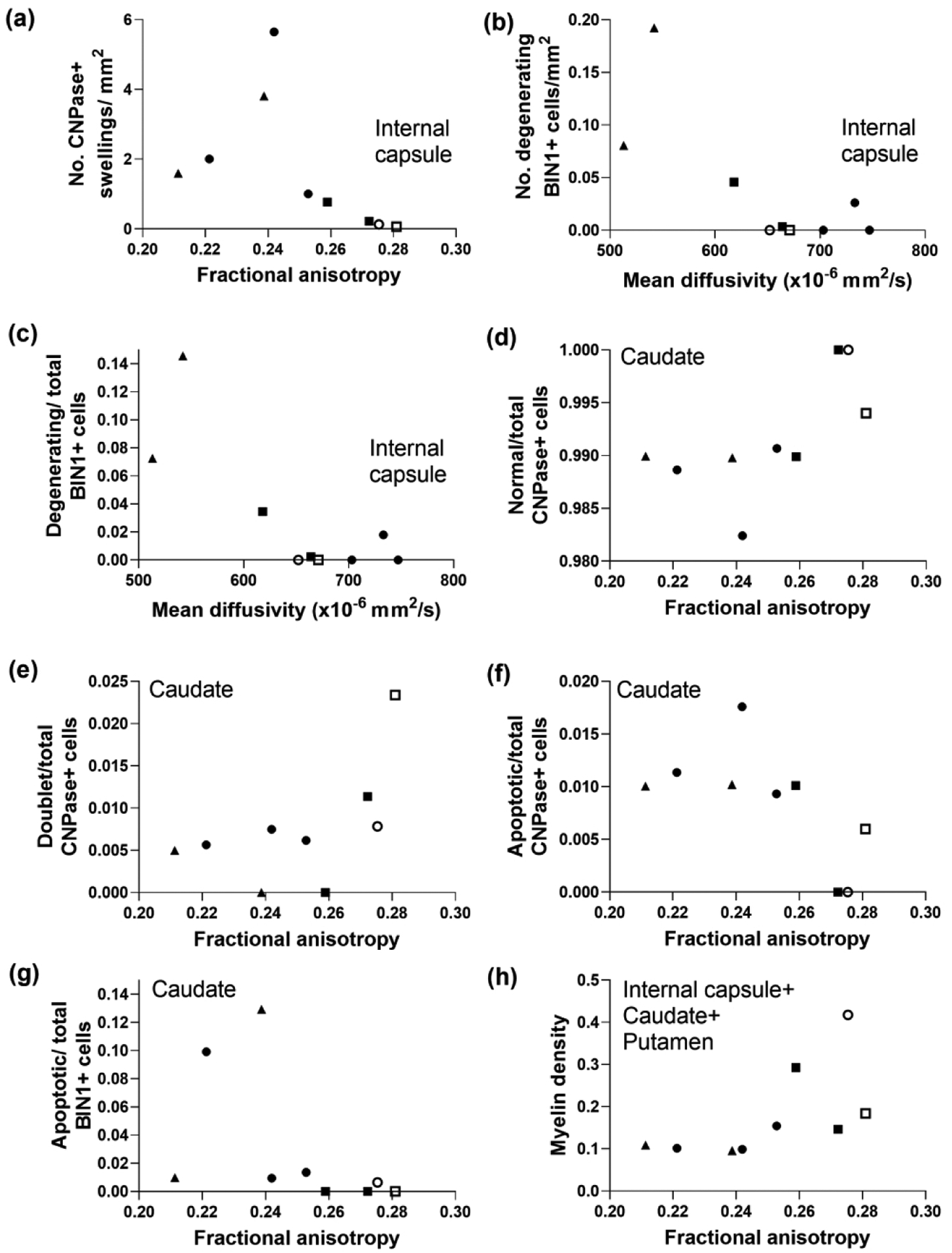
Figure 11.

Diffusion tensor imaging is associated with selective forms of oligodendrocyte degeneration and myelin injury in subacutely injured piglet striatum. (a) In the first three bundles of the internal capsule, the number of large CNPase+ myelin swellings correlated with fractional anisotropy (FA; r= −0.833, p=0.008). Mean diffusivity (MD) was associated with the number of degenerating BIN1+ cells (b; r= −0.696, p=0.046) and the ratio of degenerating BIN1+ cells to total BIN1+ cells in the internal capsule (c; r= −0.696, p=0.046). FA correlated with the ratios of normal-to-total CNPase+ cells (d; r=0.695, p=0.045), doublet-to-total CNPase+ cells (e; r=0.695, p=0.045), apoptotic-to-total CNPase+ cells (f; r= −0.695, p=0.045), and apoptotic-to-total BIN1+ cells (g; r= −0.763, p=0.024) in caudate. (h) Myelin density measured by Luxol fast blue stain in caudate, internal capsule, and putamen correlated with FA (r=0.733, p=0.031). Symbols indicate piglets that received QA (open circle: 240 nmol, solid circle: 720 nmol, and solid triangle: 960 nmol), HI (solid squares), or sham procedure (open square).
