Figure 6.

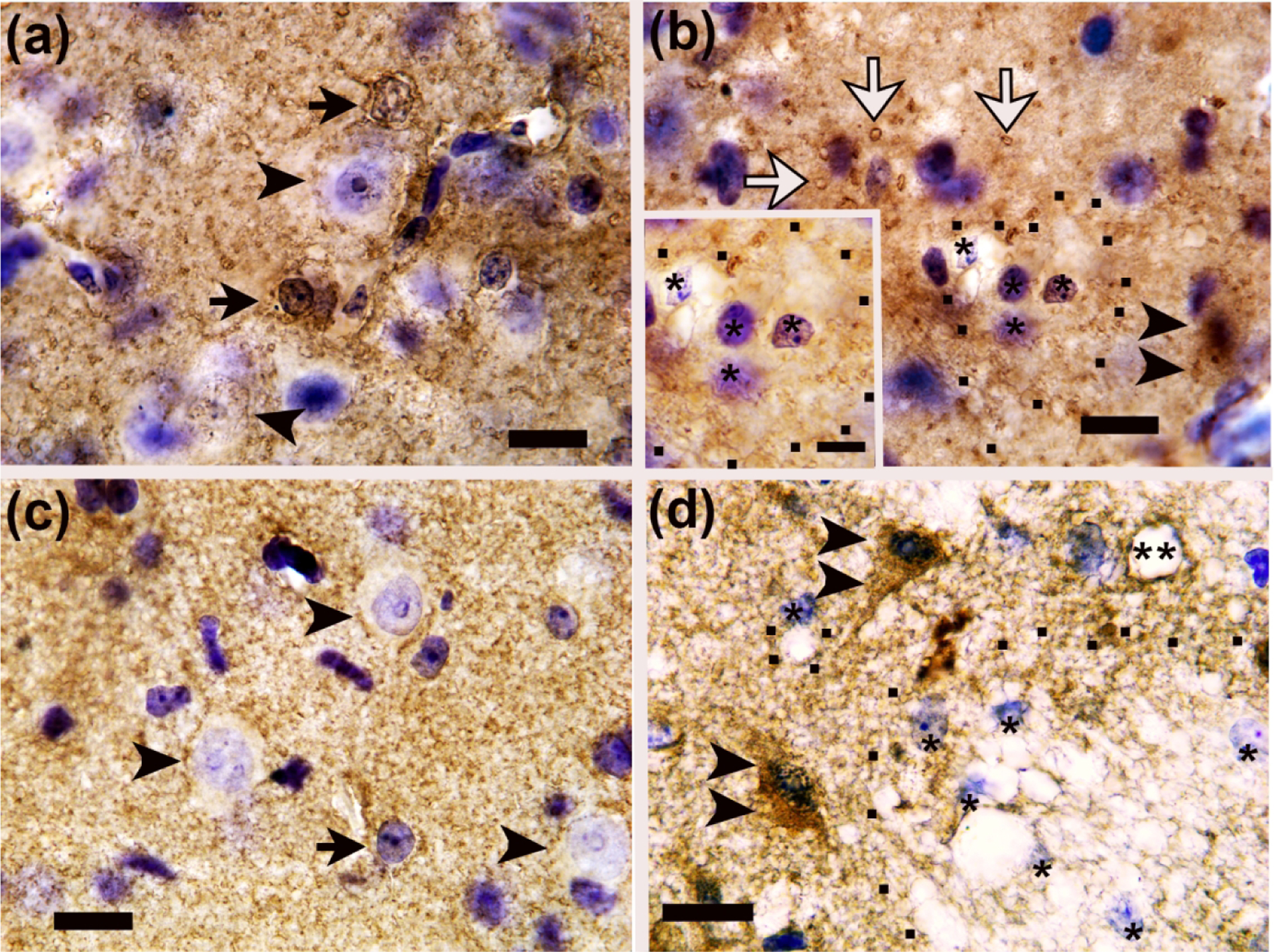
Glutamate transporter 1 (GLT-1) immunohistochemistry identifies normal and pathological astrocytes in piglets with subacute striatal injury. (a) Putamen in a sham piglet shows normal GLT-1+ astrocytes (arrows). Normal neurons have GLT-1 processes decorating their cell membranes (arrowheads). (b) After hypoxia-ischemia (HI), astrocyte swelling is apparent in the putamen. The asterisks identify nuclei of swollen GLT-1+ astrocytes, and the black dots outline the swollen astrocyte bodies. The inset shows cytoplasm vacuoles of differing sizes in swollen astrocytes. White arrows denote swollen GLT-1+ astrocyte processes. The double arrowheads show a degenerating neuron with nuclear condensation and emerging GLT-1 cytoplasmic immunoreactivity. (c) After piglets were injected with 240 nmol quinolinic acid (QA), normal GLT-1+ astrocytes (arrow) and normal neurons with GLT-1+ processes along the cell membrane (arrowheads) were visible in putamen. (d) Injection of 960 nmol QA caused astrocyte swelling in putamen. Single asterisks identify the nuclei of swollen astrocytes, and black dots outline the swollen astrocyte cell bodies. The double arrowheads show degenerating neurons with necrotic chromatin condensation in the nucleus (Martin et al., 1998), GLT-1 enrichment in the cytoplasm, and a sharply angular cell body contour. The double asterisks identify a blood vessel with perivascular astrocyte labeling in apposition to the vessel exterior without apparent vasogenic edema. Photos were taken at 1000x with oil immersion. Main panel scale bars are 10 μm. Scale bar for panel B inset is 5 μm.
