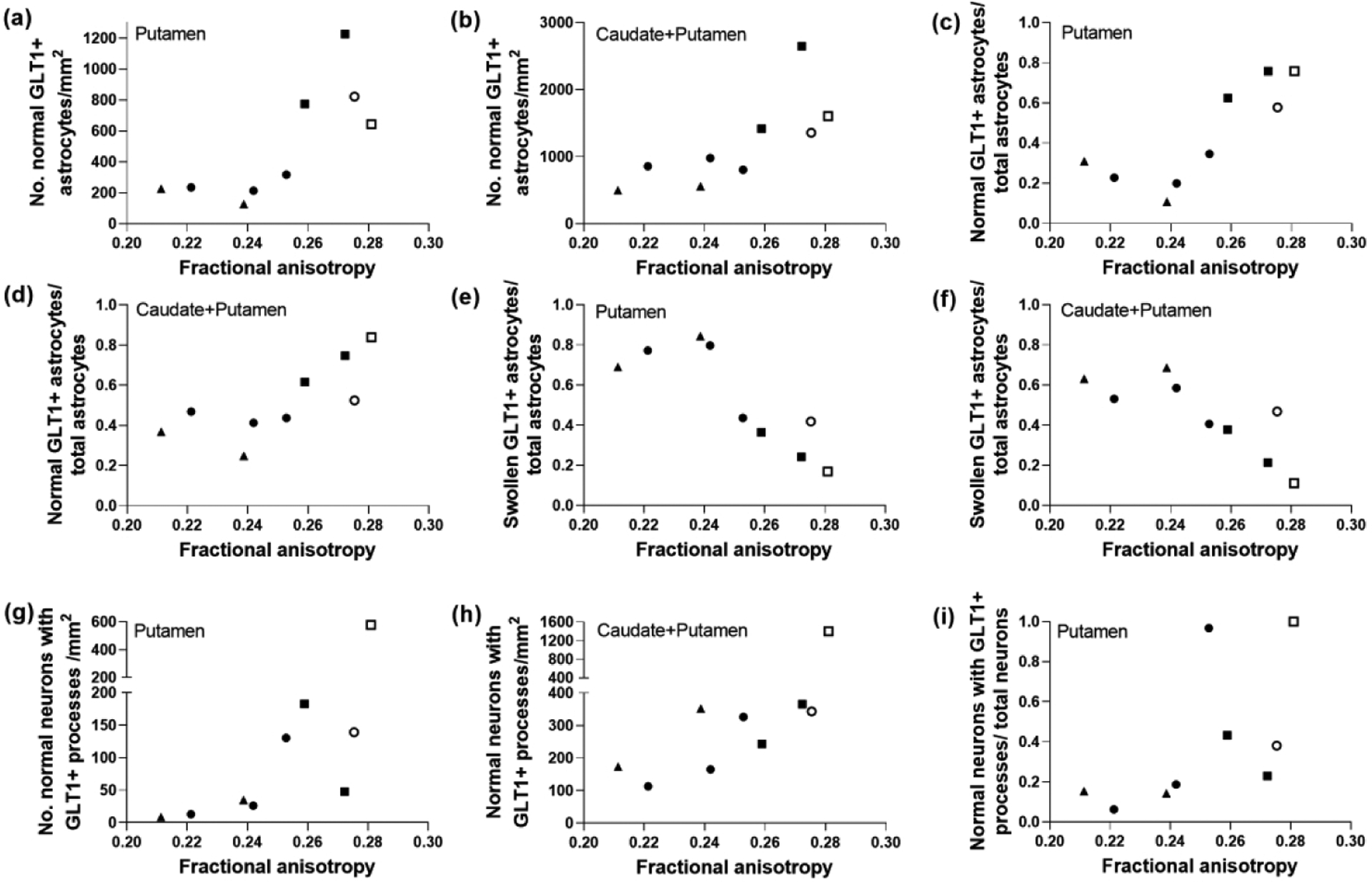
Figure 7.

Glutamate transporter 1 (GLT-1) labeling distinguished a relationship between astrocyte morphology and fractional anisotropy (FA) in piglets with excitotoxic and ischemic striatal damage. The number of normal GLT-1+ astrocytes in putamen (a; r=0.750; p=0.026) and caudate and putamen (b; r=0.833; p=0.008) correlated with FA, as did the ratio of normal GLT-1+ astrocytes to total astrocytes in putamen (c; r=0.800; p=0.014) and caudate and putamen (d; r=0.817; p=0.011). The ratio of swollen GLT-1+ astrocytes to total astrocytes also correlated with FA in putamen (e; r= −0.800; p=0.014) and caudate and putamen (f; r= −0.817; p=0.011). FA was also related to the number of normal neurons decorated by GLT-1+ processes on the neuronal cell membrane in putamen (g; r=0.900; p=0.002) and caudate and putamen (h; r=0.717; p=0.037). (i) The ratio of normal neurons with GLT-1+ processes to total neurons also correlated with FA in the putamen (r= 0.800; p=0.014). Symbols indicate piglets that received QA (open circle: 240 nmol, solid circle: 720 nmol, and solid triangle: 960 nmol), HI (solid squares), or sham procedure (open square).
