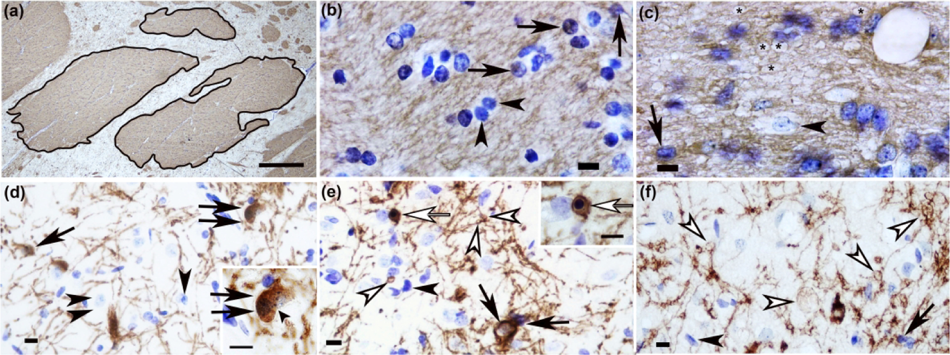
Figure 9.

CNPase immunohistochemistry identifies subacute damage to oligodendrocytes and their myelinating processes in piglet striatum with excitotoxic and ischemic injury. (a) In a coronal plane of the anterior striatum, the first three internal capsule bundles that were distinct from the corpus callosum and along the dorsal aspect of the lateral ventricle were outlined in ImageJ for area measurement. Cell counts and myelin swellings were divided by the sum area of the bundles. (b) Internal capsule of a sham piglet had normal CNPase+ oligodendrocytes with fine, delicate processes (arrows). The bundle matrix had a smooth and homogeneous meshwork of CNPase immunoreactivity. Arrowheads identify normal, CNPase-negative cells. (c) Injection of 720 nmol quinolinic acid (QA) caused myelin swellings in the internal capsule’s matrix (asterisks). The myelin swellings created a spongiform pathology that attenuated the normal, homogeneous staining typical of white matter. The arrow identifies a normal, CNPase+ oligodendrocyte. The arrowhead shows a CNPase-negative cell. (d) Caudate of a sham pig showed a CNPase+ doublet cell with two nuclei, indicative of a recently divided oligodendrocyte (double black arrows). The inset shows the doublet cell’s incompletely separated cytoplasm with two discernible nuclei. The inset’s small arrowhead shows the separation between the two nuclei. The single arrow shows a normal, CNPase+ oligodendrocyte. The main panel’s arrowhead identifies a CNPase-negative cell. The double arrowheads show a neuron. (e) After hypoxia-ischemia, oligodendrocyte process myelin swellings (white arrowheads) were apparent in putamen. An apoptotic, CNPase+ oligodendrocyte with nuclear condensation that formed a dark blue, round mass is identified by a white arrow in the main panel and inset. Normal CNPase+ oligodendrocytes (black arrows) and CNPase-negative cells (black arrowhead) are also shown. (f) Injection of 960 nmol QA led to development of oligodendrocyte process myelin swellings (white arrowheads). The arrow shows a CNPase+, normal oligodendrocyte. The black arrowhead shows a CNPase-negative cell. Panel A was photographed at 40x, and the scale bar is 0.25 mm. Panels B, C, and inset D were photographed at 1000x with oil immersion. The main panels D, E, and F were photographed at 400x. Scale bars in panels B-F are 10 μm.
