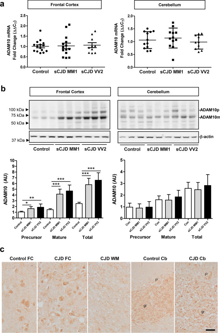
Fig. 2.
ADAM10 expression in sCJD brain tissue. a RT-qPCR analysis of ADAM10 in the frontal cortex (FC) and cerebellum (CB) of control, sCJD MM1, and sCJD VV2 cases. GAPDH was used for normalization. FC (controls n = 15, sCJD MM1; n = 11, sCJD VV2 n = 10), CB (controls n = 12, sCJD MM1; n = 12, sCJD VV2 n = 10). b Representative western blot analysis of ADAM10 in the FC and CB of control, sCJD MM1, and sCJD VV2 cases. ADAM10 precursor (ADAM10p) and mature (ADAM10m) forms are indicated. β-actin was used for normalization. Graphical representation of western blot data acquired from the analysis of FC (controls n = 12, sCJD MM1; n = 12, sCJD VV2; n = 12) and CB (controls; n = 9, sCJD MM1; n = 10, sCJD VV2; n = 10); *p < 0.05, **p < 0.01, ***p < 0.001. c ADAM10 immunohistochemistry in the frontal cortex (FC), cerebellum (Cb), and subcortical white matter (WM) in control and sCJD MM1. ADAM10 immunoreactivity is observed in neurons in the FC and granule cells (gr) of cerebellum, but Purkinje cells (P) are very weakly stained or not at all. No positive cells are distinguished in the white matter; scale bar = 20 µm

