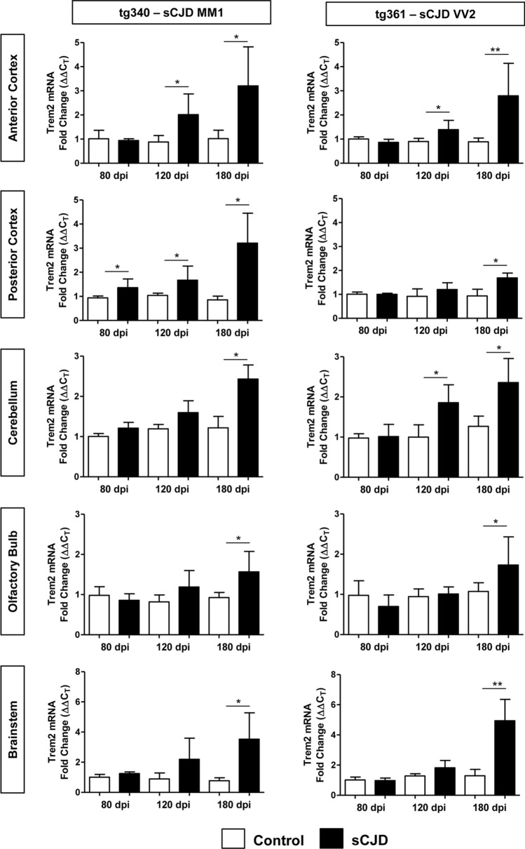
Fig. 3.
mRNA Trem2 expression in the brain of sCJD MM1 and VV2 mouse models. RT-qPCR analysis Trem2 expression in mouse models of sCJD MM1 (tg340-CJD MM1) (left panels) and sCJD VV2 (tg361-CJD VV2) (right panels) subtypes. Trem2 expression was analysed in five brain regions: anterior and posterior cortex, cerebellum, olfactory bulb, and brainstem, at three time points: pre-clinical (80 days post inoculation (dpi)), early-clinical (120 dpi), and clinical (180 dpi). CJD-inoculated animals (black columns) were compared to their corresponding control-inoculated animals (white columns). Four to eight animals per time point and condition were used. Normalization was performed using the housekeeping gene Hprt; *p < 0.05, **p < 0.01

