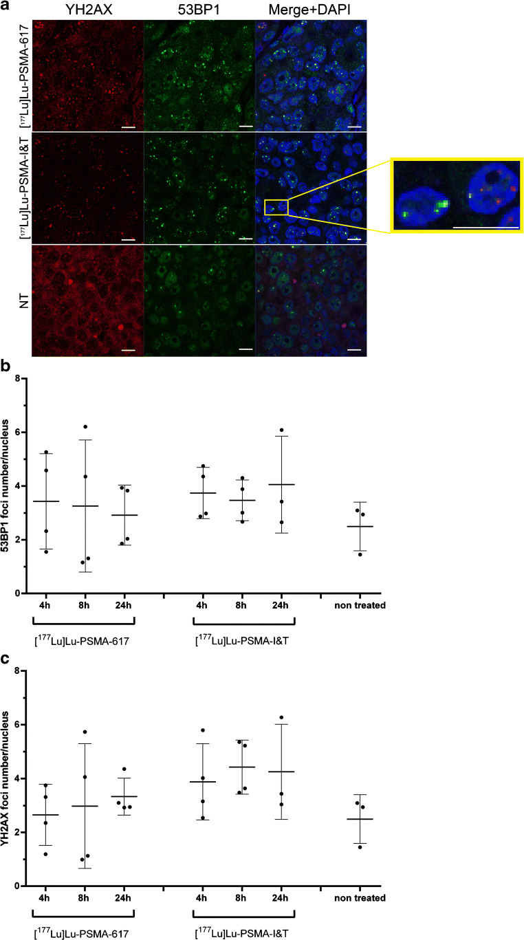
Fig. 6.
Ex vivo DNA double-strand breaks analysis on PC295 PDX tissue. a Representative images of tumor sections of mice injected with [177Lu]Lu-PSMA-617 or [177Lu]Lu-PSMA-I&T at 4 h p.i. and non-treated animals (scale bar = 10 μm). b Corresponding 53BP1γH2AX foci analysis. Average number of foci per nucleus per mouse is depicted. c Corresponding γH2AX foci analysis. Average number of foci per nucleus per mice is depicted. All error bars indicate standard deviation

