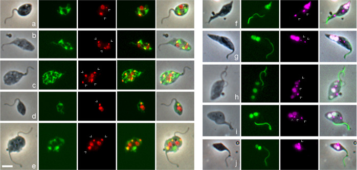
Fig. 3. Visualization of the mitochondrion and nucleus in gametes and potential meiotic intermediates.
a–e In T. b. brucei 1738 Mito GFP, GFP is targeted to the mitochondrial lumen using the 24 amino acid import signal of frataxin. L to R: phase contrast, GFP, DAPI (red), merge GFP and DAPI, merge all. DAPI fluorescence is coloured red to visualize yellow (i.e. dual red and green) fluorescence of the kinetoplast within the mitochondrion in the merged images. Kinetoplasts are indicated by arrowheads. a 2K1N gamete; b 3K2N intermediate; c 4K2N intermediate; d 1K2N intermediate; e 3K2N intermediate with two flagella. f–j T. b. brucei 1738 H2B::GFP YFP::PFR, fusion proteins visualize the nucleus and the paraflagellar rod (PFR) in the flagellum respectively. L to R: phase contrast, GFP, DAPI (magenta), merge. f 2K1N meiotic divider with very large posterior nucleus; the paler PFR belongs to the new flagellum; g 2K2N intermediate; h 3K3N intermediate; i 3K2N intermediate; j 1K2N intermediate. Scale bar = 5 µm.

