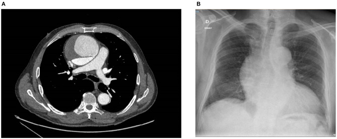
Figure 1.
(A) Contrast medium thoracic CT scan showing a CTAAD with a thick intimal tear (7 mm) and an enlarged ascending aorta (82 × 87 mm). The false lumen is partially thrombosed. (B) Chest X-ray showing an aortic aneurysm with a widening of the mediastinal silhouette, an enlargement of the aortic knob, and a displacement of the trachea from the midline. CT, computed tomography; CTAAD, chronic type A aortic dissection.

