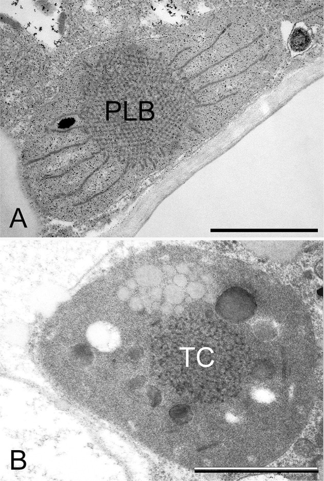
Figure 5.

Transmission electron micrographs of the prolamellar body (PLB) present in the etioplast of the cotyledon of a 2-week-old dark-germinated rosemary (Rosmarinus officinalis) seedling (A), and tubular complex (TC) of a leucoplast of the neck cell of a peltate glandular hair on the surface of a light-grown adult rosemary plant (B). Scale bar: 1 μm. Sample preparation was as described in Böszörményi et al. (2020).
