Figure 5.
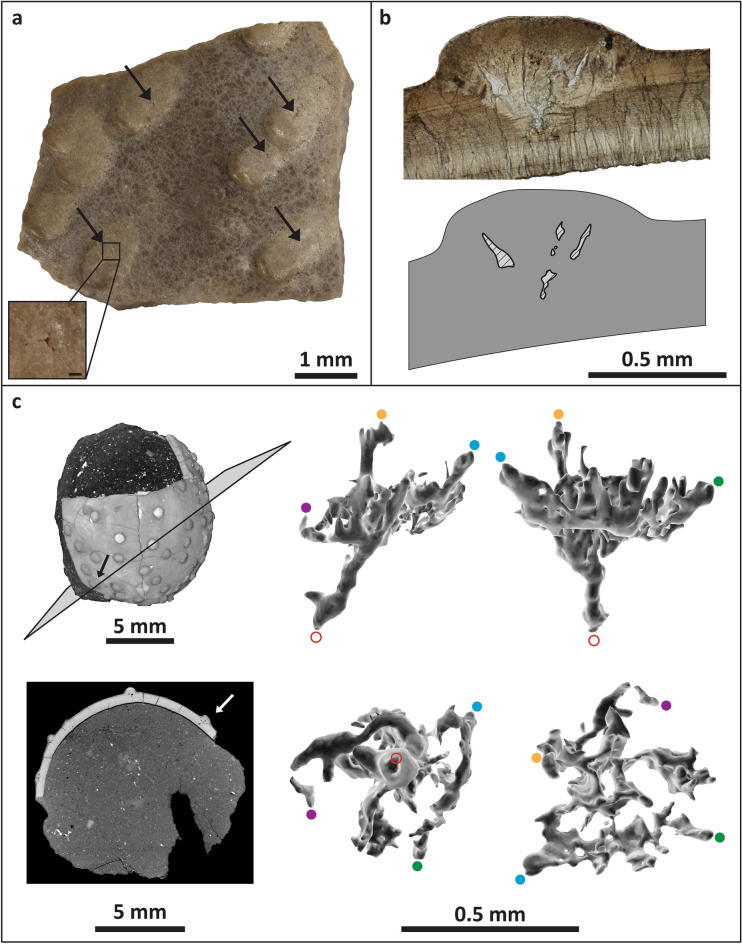
Stillatuberoolithus storrsi oogen. et oosp. nov. pore structure. (a) Outer surface of UCM 1073 showing dispersituberculate ornamentation with slightly elongated nodes. Note very small pore openings on the nodes (arrows). Inset shows pore opening with a scale of 0.2 mm; (b) Radial thin section of DMNH EPV.65602 (holotype) with diagram highlighting open cavities within the node due to branching pores; (c) Rendered CT scans of partial egg (DMNH EPV.128286, paratype). Pore canals are evident within nodes; straight, perpendicular lines in shell between nodes are fractures in the eggshell. A pore canal within a node free of cracks was selected for partitioning (arrow). Four 3D views of the selected pore canal are provided. Upper left and upper right images are lateral views. Lower left image is a view from the base of the pore canal, looking up towards the outer eggshell surface. Lower right image is a view from the top of the pore canal, looking down towards the inner surface of the eggshell. Red open circle indicates where the pore canal meets the inner surface of the eggshell. Orange and blue dots indicate where the pore reaches the outer surface of the node. Purple and green dots provided to assist with orientation. Images in (c) generated with Dragonfly software, Version 4.1.0.647 for Windows; software from Object Research Systems (ORS) Inc., Montreal, Canada is available at http://theobjects.com/dragonfly.

