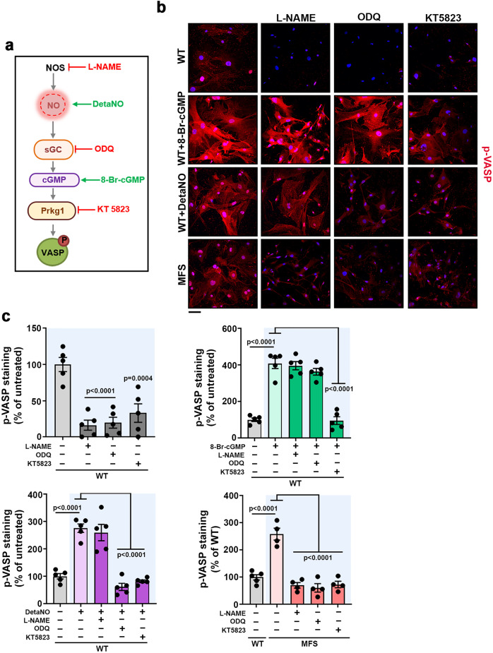
Fig. 1. Pharmacological inhibition of signaling components of the NO–sGC–PRKG pathway decreases pVASP-S239 induction in VSMCs from MFS mice.
a NO signaling components and the targets of pharmacological stimuli (green) and inhibitors (red). b Representative images of pVASP-S239 immunofluorescence (red) and DAPI-stained nuclei (blue). c Quantification of pVASP-S239 immunofluorescence in WT and MFS VSMCs treated with 300 µM L-NAME, 10 µM ODQ, or 1 µM KT5823 for 1 h before stimulation for 5 minutes with 100 µM 8-Br-cGMP or 100 µM DetaNO, as indicated. b, c Five independent cell batches were used for all conditions except for MFS VSMCs (n = 4). Scale bar, 50 μm. Data are shown relative to untreated WT cells as mean ± s.e.m. Each data point denotes the mean value from an independent experiment. Differences were analyzed by one‐way ANOVA with Dunnett’s post-hoc test (p-values are shown). Source data are provided in the Source Data file.

