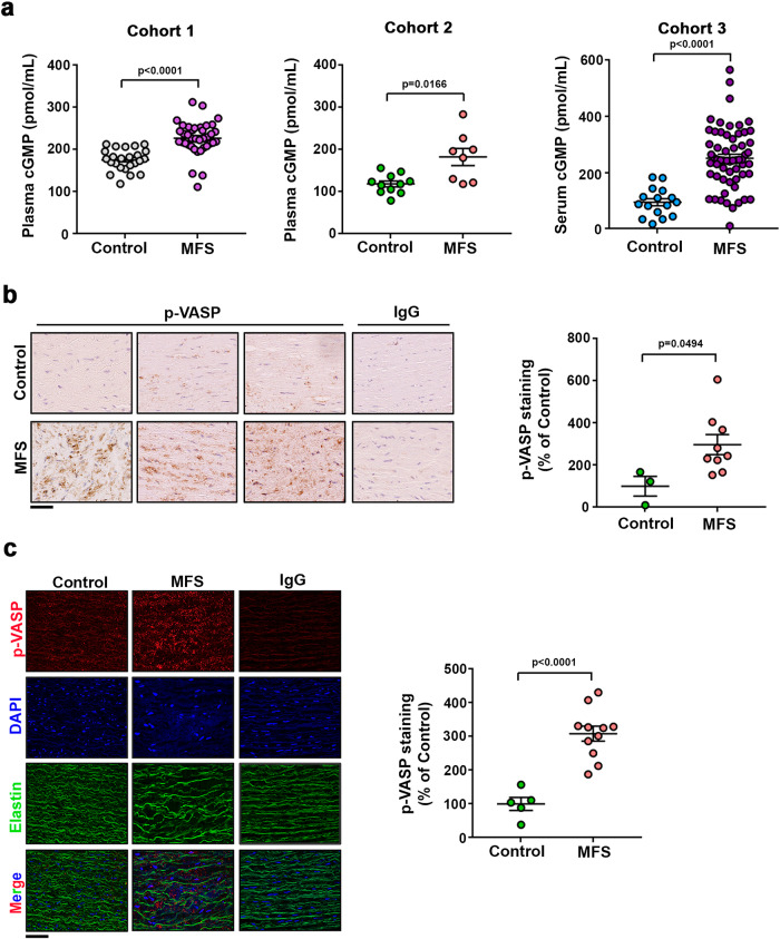
Fig. 5. The sGC–PRKG pathway is activated in aortas of MFS patients.
a Plasma (Cohorts 1 and 2) and serum (Cohort 3) cGMP in three independent cohorts including 24 healthy donors and 38 MFS patients (Cohort 1); 11 healthy donors and 8 MFS patients (Cohort 2); and 11 healthy donors and 33 MFS patients (Cohort 3). Data are mean ± s.e.m. Each data point denotes an individual. b Representative medial layer images and quantification of pVASP-S239 immunohistochemistry in aortic cross-sections of human samples from 3 control donors and 9 MFS patients. Scale bar, 50 μm. IgG staining served as a negative control. Data are shown relative to healthy donors as mean ± s.e.m. Each data point denotes an individual. c Representative medial layer images and quantification of pVASP-S239 immunofluorescence (red) and DAPI-stained nuclei (blue) in sections from 5 control donors and 11 MFS patients. Scale bars, 50 μm. IgG staining served as a negative control. Data are shown relative to healthy donors as mean ± s.e.m. Each data point denotes an individual. a–c Differences were analyzed by unpaired two-tailed t-test with Welch’s correction (p-values are shown). Source data are provided in the Source Data file.

