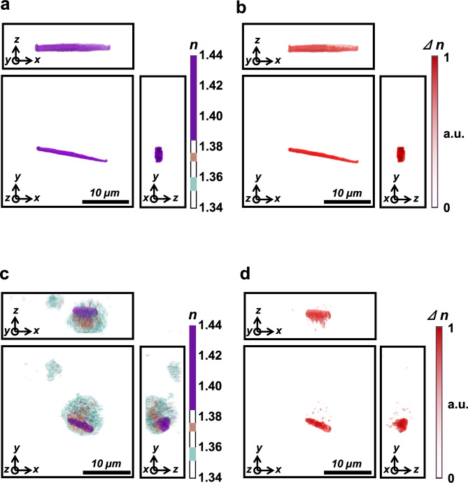
Figure 4.
Birefringence properties of patient monosodium urate (MSU) crystals and polarization-sensitive RI contrast tomogram. (a) A three-dimensional (3D) holotomographic image of synthetic MSU crystals under a polarized light source with a 0° angle. (b) Polarization contrast image of MSU crystals under a polarizing filter used during optical diffraction tomography (ODT) with a 90° angle. (c) 3D holotomographic image of synovial mononuclear cells. Intracellular MSU crystal is distinguished by its relatively higher RI value, as shown in previous figures. (d) Polarization contrast image of MSU crystal-containing synovial mononuclear cells, obtained under a polarizing filter in ODT with a 90° angle. In this image, all structures without birefringence disappeared, and the remaining MSU crystal was shown in red color. n: refractive index, ∆n: change of refractive index under the two different polarizing filter angles.

