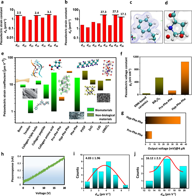
Fig. 2. Piezoelectricity of Pro-Phe-Phe and Hyp-Phe-Phe assemblies.
a, b Calculated piezoelectric strain constants for Pro-Phe-Phe (a) and Hyp-Phe-Phe (b). Selected values are marked above relevant columns. c, d Zoom-in on the electronic structure of the Hyp-Phe-Phe crystal. The π-density of the diphenylalanine motif remains identical across both crystals (c) and d shows the hydroxylated pyrrolidine structure of the Hyp residue in the Hyp-Phe-Phe crystal. e Comparison of piezoelectric strain response of different biological and non-biological materials: collagen piezoelectric response at different length scales10, 25, 53 has been emphasized as compared to that of Pro-Phe-Phe and Hyp-Phe-Phe. f The predicted voltage coefficient of Pro-Phe-Phe and Hyp-Phe-Phe in comparison with some currently used inorganic materials. g Calculated output voltage upon applied force of 4 µN. h–j Experimental measurement of coefficients using PFM. h Linear relationship between the vertical piezoresponse of Hyp-Phe-Phe as measured by the photodiode system and applied voltage. Statistical distribution of the vertical d33 coefficients (i) and shear d34 coefficients (j). Additional PFM data are given in Supplementary Figs. 5–16.

