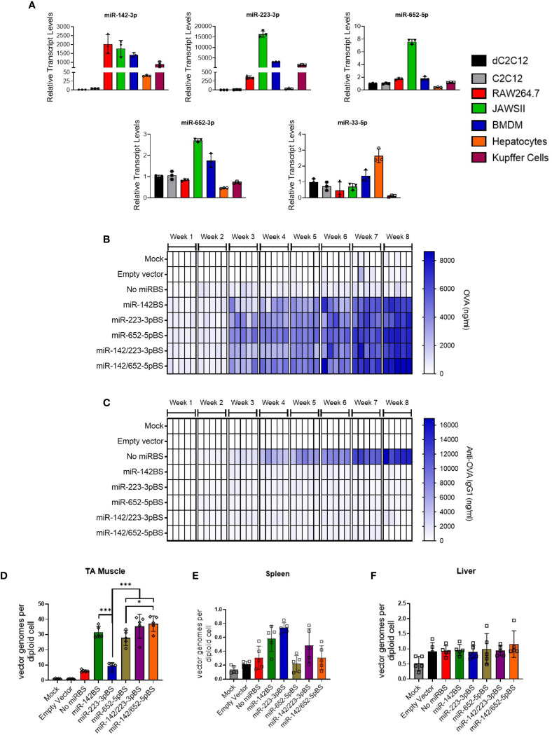
Figure 2.
Incorporation of miR-223BSs and miR-652BSs boosts in vivo OVA production and suppresses antibody development. (A) Endogenous miRNA expression levels in cultured mouse myoblasts (C2C12), myocytes (dC2C12), macrophages (RAW264.7), DCs (JAWSII), bone marrow derived macrophages (BMDM), primary mouse hepatocytes, and Kupffer cells as quantified by reverse transcription quantitative PCR (RT-qPCR) (n = 3). rAAV1 expression vectors were injected by i.m. on day 0 followed by serum collection every week for an eight-week period. (B, C) ELISA quantification of circulating OVA expression (B) and anti-OVA IgG1 (C) (1 × 10¹¹ GCs/mouse, n = 10). Single gradient heat map representing respective analyte levels (n = 5). (D–F) ddPCR detection of rAAV vector genome copies in injected skeletal muscle (D), spleen (E), and liver (F) at eight weeks post-injection (n = 5). Values represent mean ±SD. *p < 0.05, ***p < 0.001, one-way ANOVA with Tukey’s post hoc test.

