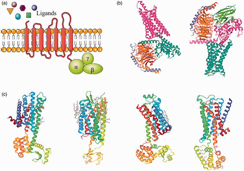
Figure 1.
Schematic diagram of a classic GPCR structure and representative three-dimensional GPCR-agonist, GPCR-antagonist complexes discussed in this review. (a) Schematic diagram of a GPCR seven-transmembrane structure. (b) Structures of the human adenosine A1 receptor-Gi2 protein complex bound to its endogenous agonist (PDB ID: 6D9H) and the adenosine A2A receptor bound to a mini-G heterotrimer (PDB ID: 6GDG). (c) Structures of the dopamine D2 receptor (PDB ID: 6CM4), P2Y1 receptor (PDB ID: 4XNV), β2 adrenergic receptor (PDB ID: 3NYA), and muscarinic M3 receptor (PDB ID: 4U15). (A color version of this figure is available in the online journal.)

