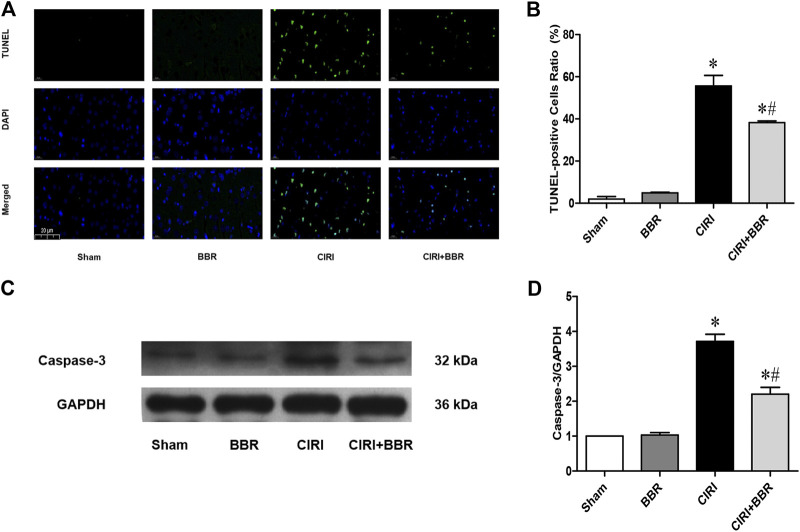
FIGURE 2.
BBR attenuates neuronal apoptosis in ischemic penumbra induced by CIRI. (A), TUNEL staining was performed on slices from cortical ischemic penumbra. (B), Ratio of positive cells. Original magnification is 400×, scale bars is 20 μm. (C), The apoptotic protein cleaved Caspase-3 from cortical ischemic penumbra was determined by Western blot. GAPDH was used as the loading control. n = 6. (D) Bar graph showing the statistical comparison of Caspase-3 protein levels across groups. Data are shown as the mean ± SEM. *p < 0.05 compared with Sham group; # p < 0.05 compared with the CIRI group.

