Figure 7.

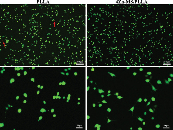
Live-dead fluorescence staining images of MG-63 cells after cultured on the poly-L-lactic acid (PLLA) and 4 zinc-doped mesoporous silica/PLLA scaffold for 1 day (dead cells indicated by red arrows).

Live-dead fluorescence staining images of MG-63 cells after cultured on the poly-L-lactic acid (PLLA) and 4 zinc-doped mesoporous silica/PLLA scaffold for 1 day (dead cells indicated by red arrows).