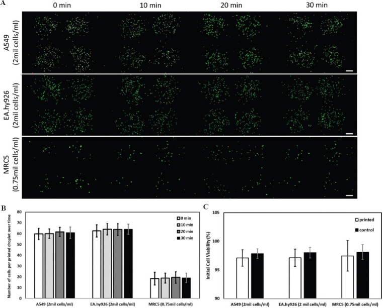
Figure 2.

(A) Representative Molecular Probes® Live/Dead stained fluorescence images of different types of printed human alveolar lung cells: A549 human lung epithelial cells (top), EA.hy926 human endothelial cells (middle) and MRC-5 human lung fibroblasts (bottom) at varying cell concentrations at different time intervals (0, 10, 20, and 30-min intervals); scale bar: 200 μm. (B) Analysis of number of printed cells per droplet over a period of 30 min (average ± standard deviation). (C) Influence of printing process on initial cell viability (Live/Dead staining kit) – printed cells versus non-printed cells (control).
