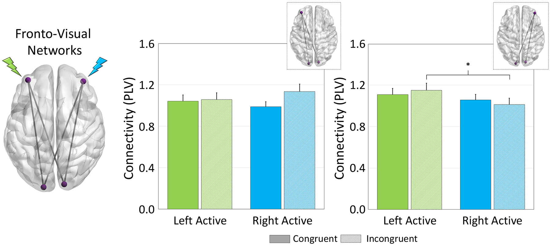
Figure 3. HD-tDCS Modulation of Fronto-Visual Networks.

(Left): The glass brain represents all functional networks interrogated (e.g., left DLPFC-left and right visual). Phase coherence among sites of stimulation during target processing were evaluated by extracting the phase-locking value (PLV; normalized to sham) and averaged over the task active period for each pathway, and then averaged across left/right occipital pathways for the left DLPFC and Right DLPFC separately. (Right) Sites of stimulation are noted in green and blue for the left- and right-DLPFC, respectively. A significant montage × condition × network interaction was apparent in the right DLPFC-bilateral visual network (far right) such that dynamic functional connectivity between right DLPFC and bilateral visual cortices was increased during processing of incongruent trials when participants underwent left DLPFC stimulation (shown in green) compared to right (p = .043). Further, there were no significant effects of tDCS configuration or task condition on left DLPFC-bilateral visual network connectivity (left graphs: p’s > .20). *p < .05
