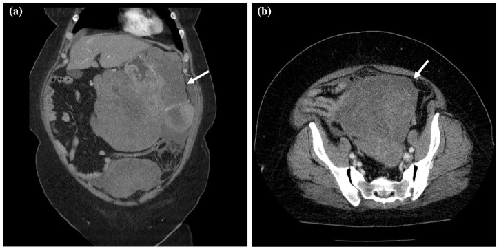
Figure 2.
CT scan images before treatment. (a) Thickening of the posterior wall of the stomach and a continuous heterogeneously enhanced mass protruding outside the stomach wall (white arrow, 23.4 × 21.1 cm). The tumor largely occupied the upper to lower left abdomen. (b) A large tumor in the pelvic cavity was also detected on the CT scan (white arrow, 13.8 × 13.1 cm).

