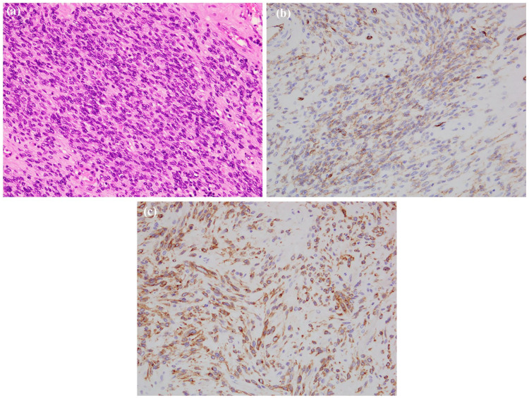
Figure 5.
Pathology of the gastric gastrointestinal stromal tumor (GIST). (a) Histology of the viable region. Spindle cells with oval nuclei arranges in fascicles. Original magnification, ×200. (b) Immunohistochemistry – tumor cells positive for CD34 and (c) immunohistochemistry – tumor cells positive for c-Kit.

