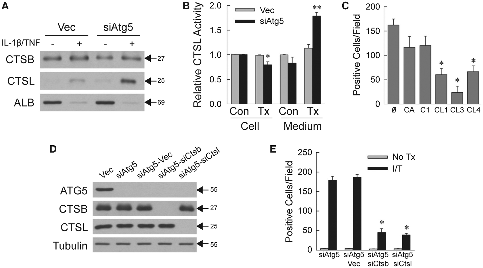
FIG. 3.

Necrosis is cathepsin-dependent. (A) Immunoblots of protein isolated from the medium of cells untreated or 24-hour IL-1β/TNF treated and probed for CTSB, CTSL, and albumin. (B) Relative CTSL activity in cells and medium from untreated control and 24-hour IL-1β/TNF-treated cells (*P < 0.01 compared with untreated Vec cells; **P < 0.000001 compared with untreated Vec cell medium; n = 5–6). (C) Numbers of trypan blue–positive siAtg5 cells that received no inhibitor or CA-074Me, Cathepsin Inhibitor I, Cathepsin L Inhibitor I, Cathepsin L Inhibitor III, or Cathepsin L Inhibitor IV for 1 hour before IL-1β/TNF (*P < 0.001 compared with no inhibitor; n = 4–6). (D) Immunoblots of total protein from the indicated cells. (E) Numbers of trypan blue–positive cells in untreated and IL-1β/TNF-treated cells at 24 hours (*P < 0.0000001 compared with IL-1β/TNF-treated siAtg5-Vec cells; n = 6–12). Molecular weights (in kD) are indicated by arrows on western blots, which are representative of three independent experiments. Abbreviations: Ø, no inhibitor; ALB, albumin; C1, Cathepsin Inhibitor I; CA, CA-074Me; CL1, Cathepsin L Inhibitor I; CL3, Cathepsin L Inhibitor III; CL4, Cathepsin L Inhibitor IV; I/T, IL-1β/TNF-treated; No Tx, untreated; Tx, IL-1β/TNF-treated.
