Figure 4.

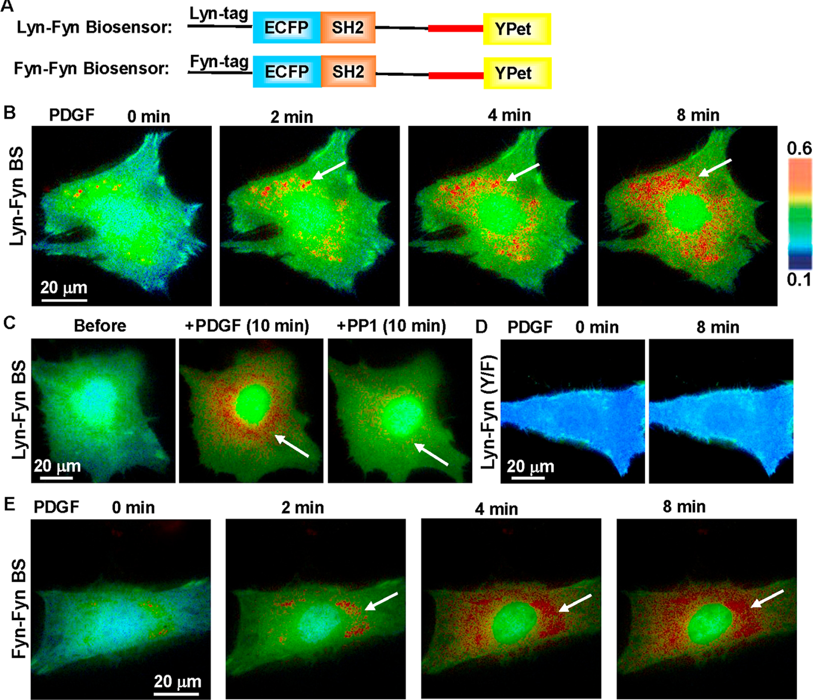
Detection of Fyn activity around the perinuclear area by the membrane-targeted biosensor. (A) The schematics of Lyn- and Fyn N-terminal peptide-tagged (Fyn-Fyn) biosensors. (B) The ratiometric (ECFP/FRET) images of the Lyn-Fyn biosensor in a representative MEF before and after PDGF stimulation. The arrows point to the perinuclear regions of high FRET activity. (C) The high FRET of the Lyn-Fyn biosensor around the perinuclear area was inhibited by Src family inhibitor PP1 (10 μM). (D) The Y/F mutant of the Lyn-Fyn biosensor displays low and relatively uniform FRET distribution in MEF, distinct from the wild-type biosensor. (E) The Fyn-tagged Fyn biosensor displays a similar FRET distribution pattern as the Lyn-Fyn biosensor, with high FRET activity at perinuclear regions (arrows) in MEFs.
