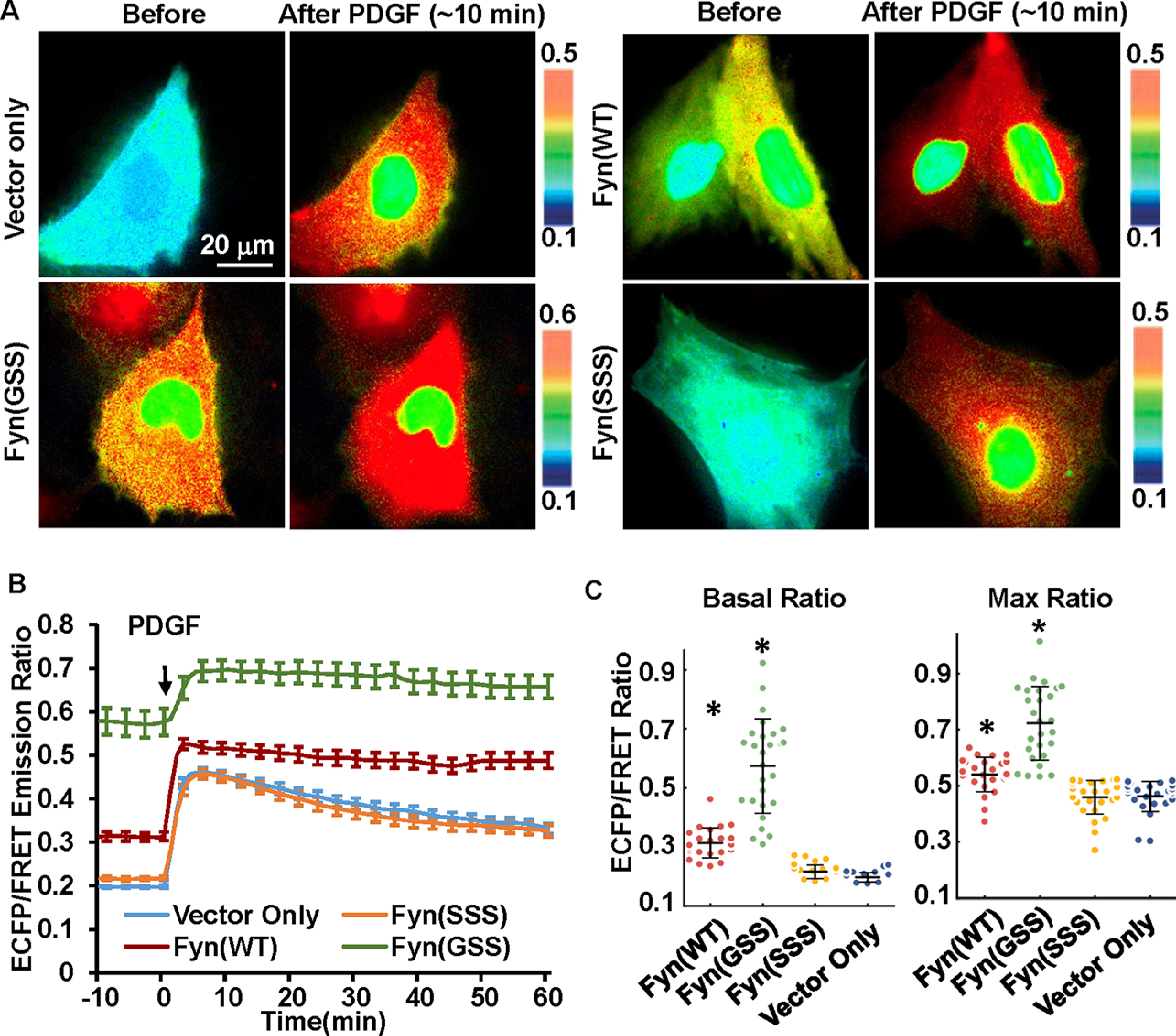
Figure 6.

The effect of Fyn N-terminal fatty acylation on Fyn activation in MEF cells. Change of palmitoylation or myristoylation modification at the Fyn N-terminal was introduced by mutation of wild-type Fyn(GCC) to Fyn(GSS) or Fyn(SSS). (A) Representative ECFP/FRET ratio images of PDGF-stimulated MEF cells cotransfected with the cytosolic Fyn biosensor (1.0 μg of DNA on a 24-well plate) with vector only or the indicated Fyn mutants (0.3 μg of DNA each). (B) The average time courses of the ECFP/FRET ratio of the cytosolic Fyn biosensor in cells coexpressing the Fyn mutants or control vector in (A) when they are treated with PDGF (mean ± SEM). (C) The scatter plots with mean ± SD compare the basal level and maximal ECFP/FRET ratio in cells with time courses shown in (B). (* indicates a statistically significant difference when compared with the control vector group by Student’s t test, p < 1.0e-4, n = 25, 27, 29, 27). In cells cotransfected with Fyn(WT), Fyn(GSS), Fyn(SSS), or the control vector, the ECFP/FRET ratio values (mean ± SD) were, respectively, 0.31 ± 0.05, 0.57 ± 0.16, 0.22 ± 0.023, or 0.20 ± 0.016 at the basal level and 0.54 ± 0.06, 0.72 ± 0.13, 0.46 ± 0.06, or 0.46 ± 0.05 at peak after PDGF stimulation.
