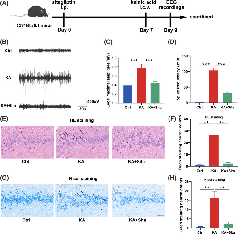
Fig. 1.
Inhibition of DPP4 attenuates KA-induced epilepsy. a Flowchart of the experiment in vivo. b Representative EEG traces of electrographic activity in control (n = 6), kainic acid (n = 9), and kainic acid + sitagliptin (n = 10) groups. c The local maximum amplitude and d spike frequency in EEG data were calculated by LabChart 8 software. e, g Representative HE and Nissl staining of the hippocampal region. Black arrows indicate damaged neurons. Scale bar = 40 μm. f, h The number of damaged neurons for each group was counted at high magnification (n = 5–6 per group). Data are presented as the mean ± SEM. **P < 0.01, ***P < 0.001

