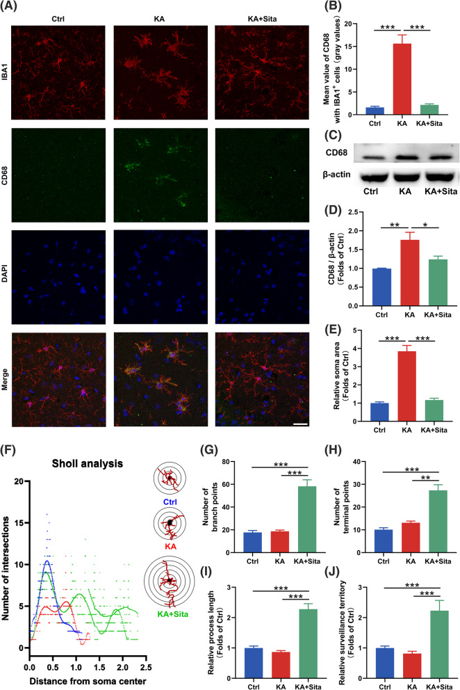
Fig. 3.
Inhibition of DPP4 alters the activation state of microglia in epileptic mice. a Representative overlay images of IBA1/CD68/DAPI staining in the hippocampus of the three groups (IBA1, red; CD68, green; DAPI, blue). Scale bar = 25 μm. b The statistical analysis of CD68 mean value in IBA1+ microglia. c, d Western blotting for CD68 in the hippocampus. Relative CD68 expression was normalized to β-actin (n = 3). e The soma area, f Sholl intersections, g process branch points, h terminal points and i the relative length of processes of IBA1+ microglia in the hippocampus of the three groups (n = 12–18 cells in 3 mice per group). j The average area covered by individual IBA1+ microglia in each group were calculated by LAS X software (n = 12–18 cells in 3 mice per group). Data are presented as the mean ± SEM. **P < 0.01, ***P < 0.001

