Figure 2.

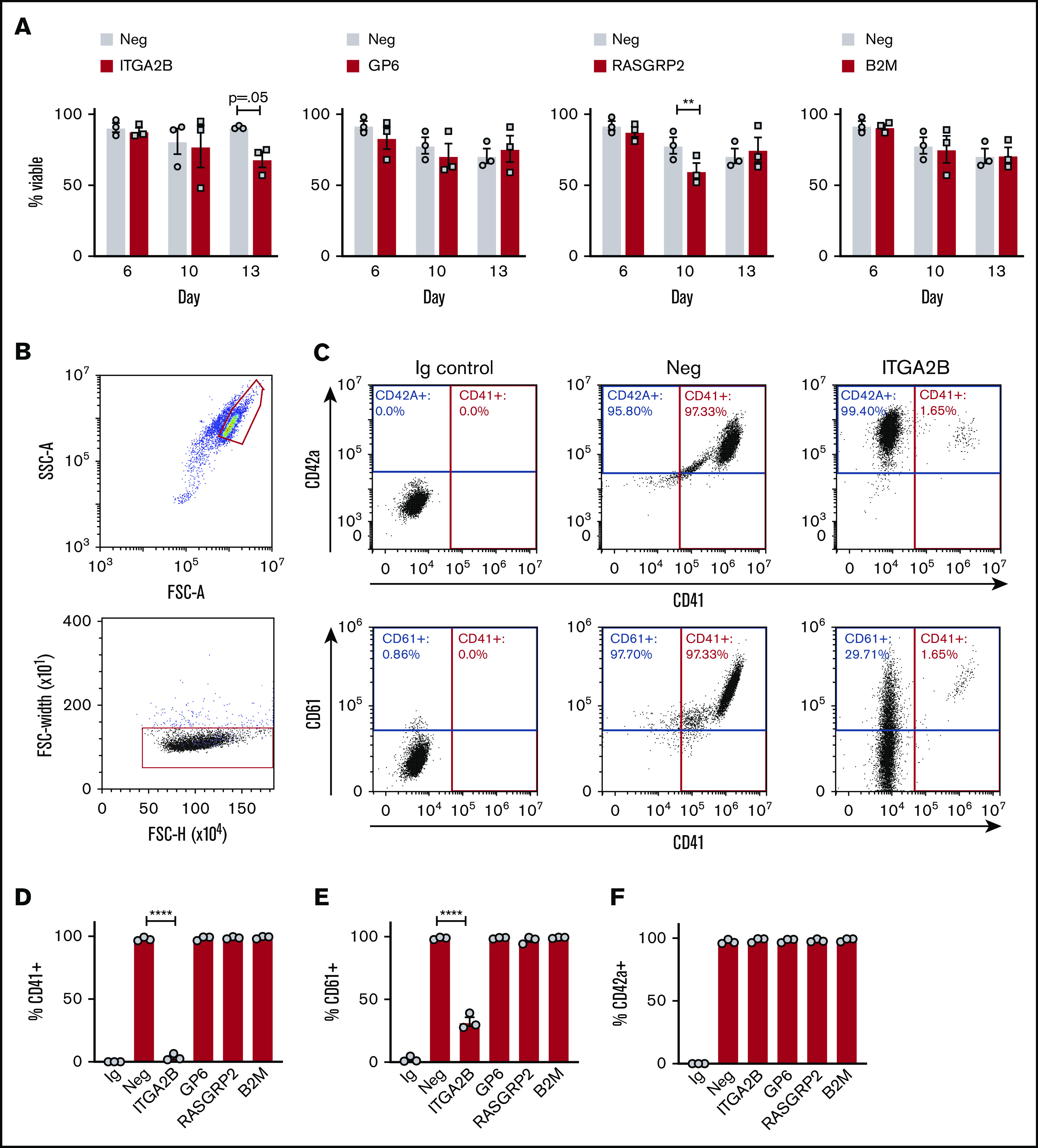
CRISPR KO cells differentiate into MKs. (A) Percent viable negative control and KO cells on days 6 to 13 of culture after CRISPR/Cas9 on day 5 (3 independent cords per group). (B) Representative flow cytometry gating strategy for MKs on day 13 of culture: single MKs were first gated on larger, lower granularity cells according to forward scatter (FSC-A) and side scatter (SSC-A) as previously published6 and further gated against doublets (FSC-H [height] vs FSC-W [width]). (C) Representative flow cytometry analysis of MK maturation markers on negative control and ITGA2B CRISPR MKs. MKs were gated as in panel B, and positive gates for MK maturation markers CD41 (x-axis), CD42a (y-axis, top panels), or CD61 (y-axis, bottom panels) were set with reference to isotype (immunoglobulin [Ig]) controls. (D-F) Mean ± SEM of the percent of cells expressing MK markers (y-axis) after deletion with CRISPRs targeting the genes listed on the x-axis (3 independent cords per group). Mixed effects analysis with Dunnett’s adjustment for multiple comparisons. Data are presented as mean ± SEM. Paired Student t tests: **P < .01; ****P < .0001.
