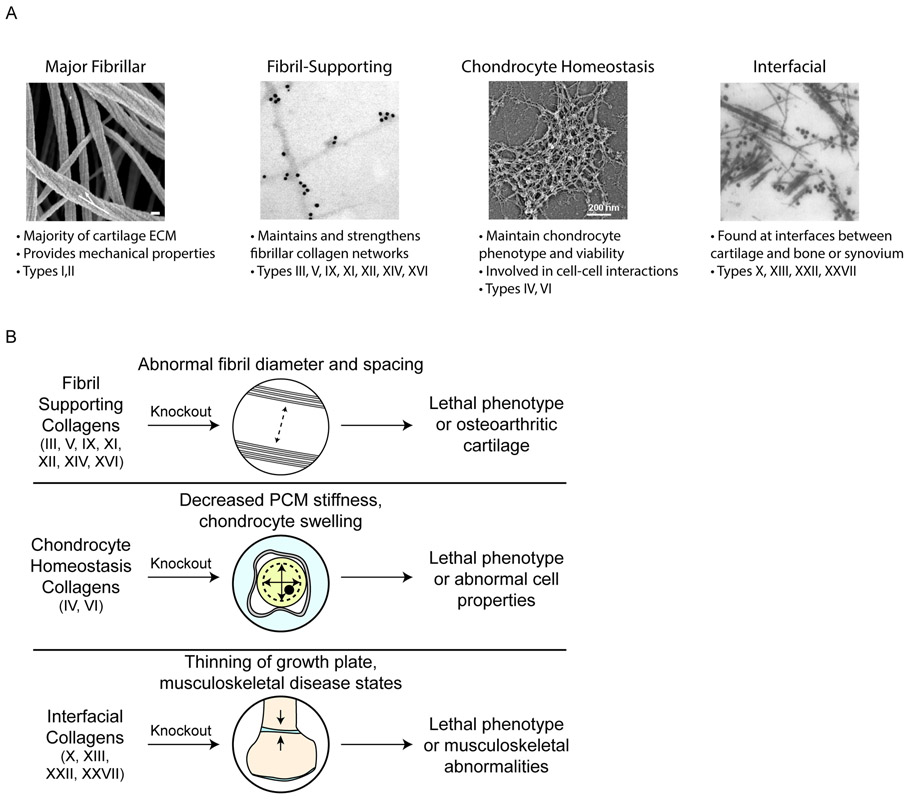
Figure 5. Functional groups of collagen subtypes in cartilage.
a ∣ (left to right) SEM image of collagen fibres in knee articular cartilage (white arrow: twisting of fibrils in the axial direction; scale bar, 100 nm); immunogold EM image of labelled collagen IX; rotary shadowing EM showing a collagen type VI network; and immunogold EM image of labelled collagen type X. b ∣ Knocking out different categories of minor collagens can cause lethal or abnormal phenotypes in cartilage and other musculoskeletal tissues. SEM, scanning electron microscopy; EM, electron microscopy. PCM, pericellular matrix. In panel a, the images (left to right) are reproduced with permission from ref. 168, Public Library of Science; ref. 249, American Society for Microbiology; ref. 250, American Society for Biochemistry and Molecular Biology; and ref. 251. Elsevier.

