
Figure 1.
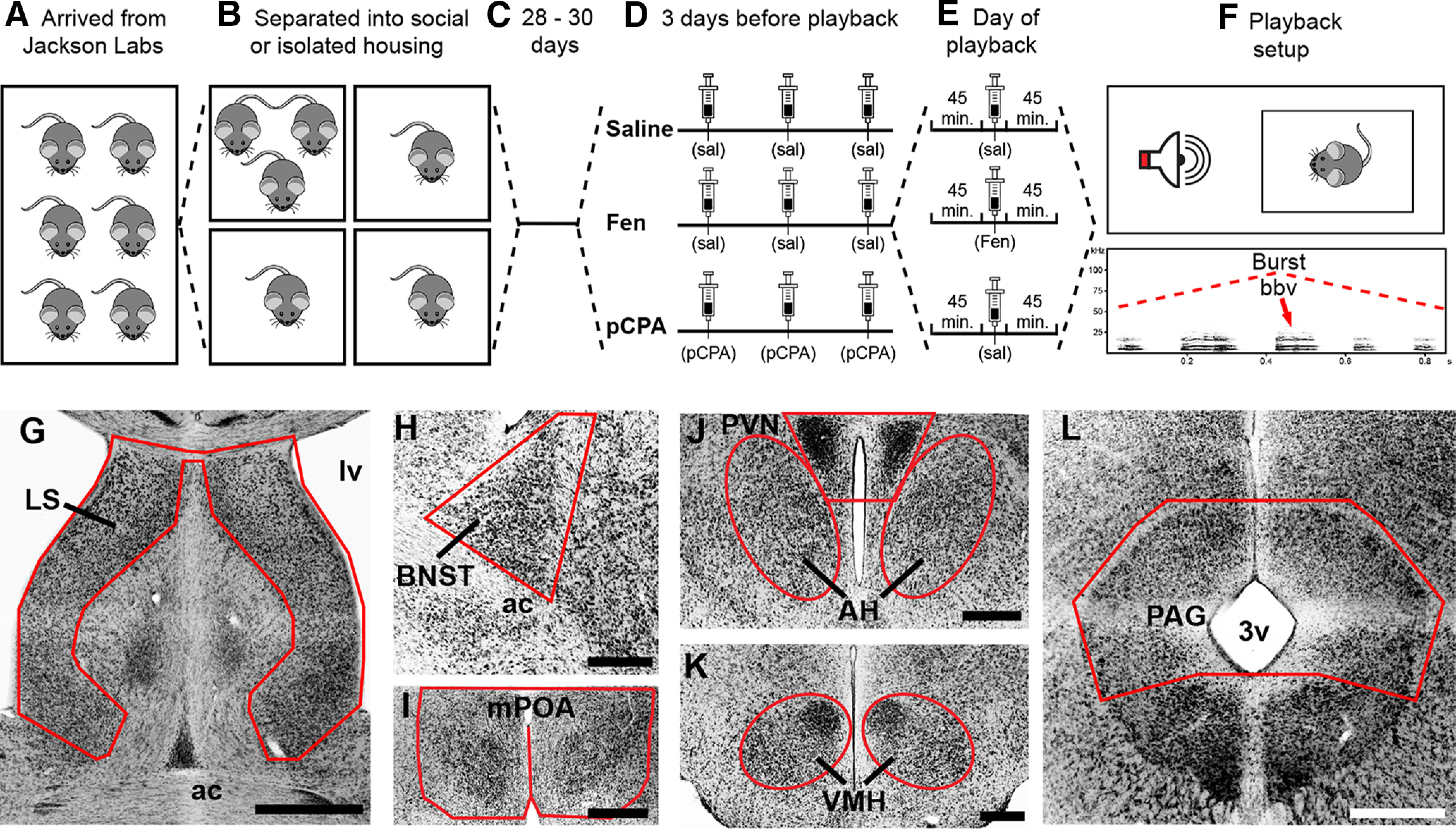
Experimental design: playback paradigm and neuroanatomy. A, Male CBA/J mice arrived from The Jackson Laboratory at 18–24 d postnatal and were immediately separated into social (three per cage) or isolated (one per cage) housing (B). C, Mice remained in their respective housing conditions for 28–30 d. D, SAL and FEN mice received SAL injections for 3 d before playback; mice in the pCPA group received pCPA injections on these days. E, Forty-five minutes before playback trials, mice in the SAL and pCPA group received SAL injections, whereas FEN mice received FEN. F, Playback trials were 60 min and consisted of 14–15 bursts of five female BBVs. G–L, Representative inverse fluorescent 10× photomicrographs showing seven nodes of the SBN: LS (G), BNST (H), mPOA (I), PVN and AH (J), VMH (K), and PAG (L). ac, anterior commissure; lv, lateral ventricle; 3v, third ventricle. Scale bars: 1 mm (G) and 500 μm (H–L).