Fig. 3.
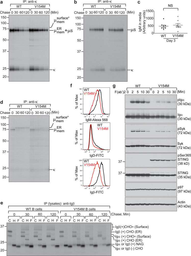
LPS-stimulated plasmablasts from STING V154M mice degraded the BCR rapidly, resulting in a significant decrease in the BCR expression on the cell surface and reduced BCR signaling upon activation. B cells from WT and V154M mice were stimulated with LPS for 3 days, starved in cysteine- and methionine-free medium for 1 h, radiolabeled for 15 min, and chased for the indicated times. Intracellular and extracellular IgM were immunoprecipitated from lysates (a) and culture medium (b), respectively, using an anti-κ antibody. The immunoprecipitates were analyzed by SDS-PAGE and autoradiography. The asterisk denotes endo‐H‐resistant complex glycans. c B cells purified from the spleens of WT (n = 8) and V154M (n = 8) mice were cultured at 106 cells per mL in RPMI medium containing LPS (20 μg/mL) for 3 days. The levels of secretory IgM in the medium were determined by ELISA (means ± SEM). d B cells from WT and V154M mice were stimulated with LPS for 3 days, starved in cysteine- and methionine-free medium for 1 h, radiolabeled for 15 min, and chased for the indicated times. The cells were lysed in Triton X‐114, and the lysates were subjected to phase separation. Intracellular membrane‐bound IgM was immunoprecipitated from Triton X‐114‐associated protein fractions using an anti‐κ antibody. The asterisk denotes endo‐H‐resistant complex glycans. e B cells from WT and V154M mice were stimulated with LPS for 3 days, starved in cysteine- and methionine-free medium for 1 h, radiolabeled for 15 min, and chased for the indicated times. Lysates were immunoprecipitated with an anti-Igβ antibody. Immunoprecipitated Igα/Igβ heterodimers were eluted from the beads and treated with endo-H or PNGase F before analysis by SDS-PAGE and autoradiography. CHO, CHO*, and NAG indicate high mannose-type glycans, complex-type glycans and N-acetylglucosamines, respectively. f B cells from WT and V154M mice were stimulated with LPS for 3 days and surface stained with IgM-Alexa 568, with B220-BV605 and IgD-FITC, or with B220-BV605 and Igβ-FITC. Gated live cells were analyzed for the expression of IgM (upper panel); gated live B220+ populations were analyzed for the expression of IgD (middle panel); and gated live B220+ populations were analyzed for the expression of Igβ (lower panel). g B cells from WT and V154M mice were stimulated with LPS for 3 days, activated with goat anti-mouse IgM F(ab’)2 (20 μg/mL) for the indicated times and lysed for immunoblot analyses of the indicated proteins

