Fig. 6.
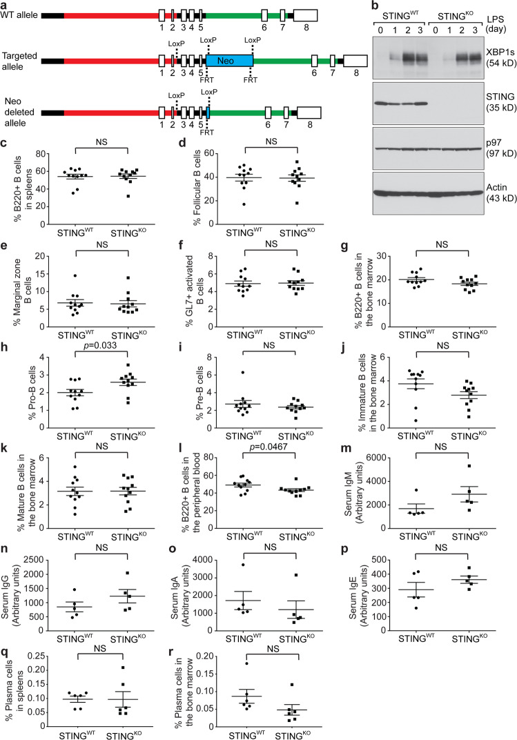
B cell-specific STINGKO mice exhibited nearly normal B cell compartments and serum antibody titers. a WT, targeted and Neo-deleted alleles are shown. Exons 3-5 of the STING gene were flanked by LoxP sites. The Neo cassette flanked by FRT sites was removed by mating chimeras carrying the successfully targeted allele with C57BL/6 FLP mice. The resultant mice carrying the Neo-deleted allele were mated with CD19Cre mice to generate B cell-specific STINGKO mice. b Purified STINGWT and STINGKO B cells were stimulated with LPS (20 μg/mL) for 3 days. Lysates were immunoblotted for the indicated proteins. Quantification of B220+ B cells (c), follicular B cells (d), marginal zone B cells (e), and GL7+ activated B cells (f) in the spleens of unimmunized STINGWT (n = 11) and STINGKO (n = 11) mice. Quantification of total B cell progenitors (g), pro-B cells (h), pre-B cells (i), immature B cells (j), and mature B cells (k) in the bone marrow of unimmunized STINGWT (n = 11) and STINGKO (n = 11) mice. l Quantification of B220+ B cells in the peripheral blood of unimmunized STINGWT (n = 11) and STINGKO (n = 11) mice. Serum levels of total IgM (m), IgG (n), IgA (o) and IgE (p) in unimmunized STINGWT (n = 5) and STINGKO (n = 5) mice were determined by ELISA. Quantification of CD138+/XBP1s+ plasma cells in the spleens (q) and bone marrow (r) of unimmunized STINGWT (n = 6) and STINGKO (n = 6) mice

