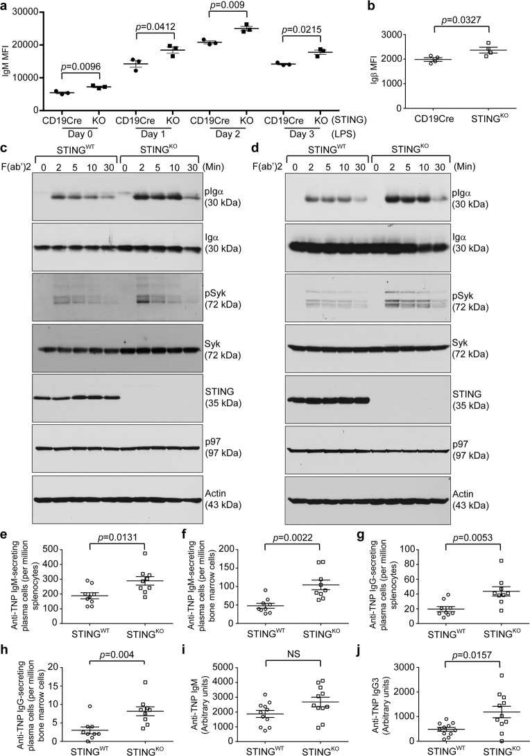
Fig. 7.
Freshly purified B cells, LPS-stimulated plasmablasts, and plasma cells from B cell-specific STINGKO mice exhibited significantly higher levels of surface BCR and were more responsive to BCR activation than those from STING-proficient mice; TNP-Ficoll-immunized B cell-specific STINGKO mice generated significantly more TNP-specific plasma cells and antibodies than TNP-Ficoll-immunized STINGWT mice. a Freshly purified B cells from CD19Cre (n = 3) and B cell-specific STINGKO (CD19Cre/STINGflox/flox; n = 3) mice were stimulated with LPS (20 μg/mL) for 3 days. Each day, cells were surface stained with B220-BV605 and IgM-PE-Cy7. Gated B220+ populations were analyzed for the expression of IgM. The mean fluorescence intensity (MFI) of IgM was plotted as the mean ± SEM. b CD19Cre (n = 4) and B cell-specific STINGKO (n = 4) mice were intraperitoneally immunized with TNP-Ficoll on Day 0. On Day 9, bone marrow cells from immunized mice were stained with B220-Alexa 488, CD138-PE, and Igβ-APC. Gated B220–/CD138+ plasma cells were analyzed for the expression of Igβ. The MFI of Igβ was plotted as the mean ± SEM. c Freshly purified B cells from STINGWT and STINGKO mice and (d) those B cells stimulated with LPS for 3 days were activated with goat anti-mouse IgM F(ab’)2 (20 μg/mL) for the indicated times and lysed for immunoblot analysis. e-j STINGWT and B cell-specific STINGKO mice were intraperitoneally immunized with TNP-Ficoll on Day 0. Immunized mice were sacrificed on Day 9, and the presence of TNP-specific plasma cells in the spleens and bone marrow was analyzed. Anti-TNP IgM-secreting plasma cells in the spleens (e) and bone marrow (f) of TNP-Ficoll-immunized STINGWT (n = 9) and B cell-specific STINGKO (n = 9) mice were quantified by ELISPOT. Anti-TNP IgG-secreting plasma cells in the spleens (g) and bone marrow (h) of TNP-Ficoll-immunized STINGWT (n = 9) and B cell-specific STINGKO (n = 9) mice were quantified by ELISPOT. Nine days after the single immunization, serum levels of anti-TNP IgM (i) and IgG3 (j) in TNP-Ficoll-immunized STINGWT (n = 11) and B cell-specific STINGKO (n = 11) mice were determined by ELISA

