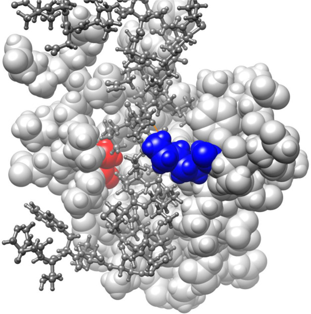
Fig. 3. 3D model to demonstrate predicted consequences of SIN3A missense variant in our cohort.

This 3D-model is based on the solution structure of mouse Sin3A PAH1 bound to the Sin3 interaction domain (SID) of SAP25 (Sin3A Associated Protein 25) displayed using UCSF Chimera v1.14 (Pattersen et al. 2004). The PAH1 domain of Sin3A (residues 119-189; sphere model, light grey) with residues Ala126 (red) and Lys155 (blue) highlighted. The SAP25 protein SID domain (residues 126-186; ball and stick model, dark grey) binds in the fold formed by the four helices of Sin3A PAH1. Sin3A Lys155 is predicted to form a 2.2A hydrogen bond with the polar side chain of Gln143 of SAP25 (orange line).
