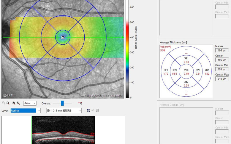
Fig. 1. The standard macula protocol of spectral domain optical coherence tomography (Spectralis, Heidelberg, Germany; software version 6.0) was used.
Early Treatment Diabetic Retinopathy Study (ETDRS) circle is the retinal thickness map analysis of the different layers for each of seven areas. The circle consists of three rings; 1-mm, 3-mm, and 6-mm diameter at the fovea. The inner and outer rings were then divided into four zones: superior, nasal, inferior, and temporal. Global macular volume for each layer also was recorded. Layer-by-layer segmentation was done automatically by using the new software for the Spectralis OCT. The following macular measurements were described; inner retinal layers, mRNFL, mGCL, mIPL, macular inner nuclear layer, macular outer plexiform layer, macular outer nuclear layer, photoreceptors, and retina pigment epithelium.

