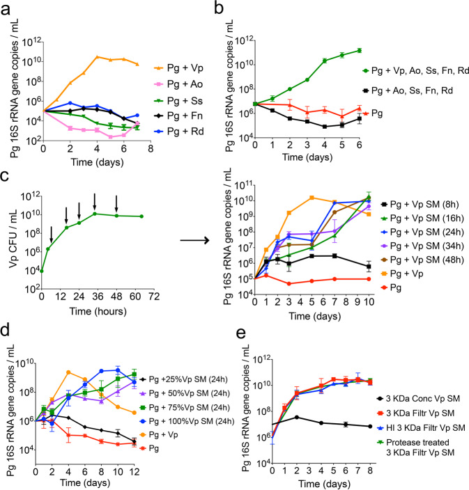
Fig. 3. V. parvula (Vp) is the key species that through a diffusible factor supports growth of low-cell-density P. gingivalis (Pg).
a Growth of Pg when co-inoculated (at 105 cells mL−1) in mucin-serum with either Vp, Actinomyces oris (Ao), Streptococcus sanguinis (Ss), Fusobacterium nucleatum (Fn) or Rothia dentocariosa (Rd). Graph shows Pg growth as determined via qPCR. b Presence of Vp is essential for the growth of a low cell-density inoculum of Pg. Graph shows Pg growth, as determined via qPCR, when inoculated together with all initial colonizers, in the absence of Vp, or as a monoculture. c Evaluation of the effect of Vp spent medium (SM), collected at different times during Vp growth, on growth of low cell-density Pg. SM was collected from a Vp batch culture grown in mucin-serum. Green curve in left panel indicates Vp cell concentrations during growth and arrows show times at which Vp SM was collected. Right panel shows growth of low cell-density Pg (105 cells mL−1) in Vp SM collected at different time points of the Vp growth curve. d Evaluation of the effect of different concentrations of Vp SM (collected at 24 h) on growth of a low-cell-density Pg inoculum showing threshold-dependent stimulation of growth by Vp SM. e Soluble factor in Vp SM capable of supporting growth of low-cell-density Pg is smaller than 3 kDa, is heat-stable and is protease resistant. SM from Vp grown for 24 h in mucin-serum was filtered through 3 kDa membranes and either heat-inactivated (HI) or treated with proteases, followed by lyophilization and reconstitution (10x) in dIH20. Reconstituted fractions (Conc = >3 kDa and Filtr = <3 kDa) were added to fresh mucin-serum medium (1:3, vol:vol) to evaluate growth of low-cell-density Pg (105 cells mL−1).

