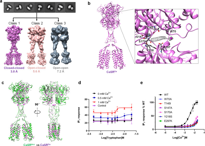
Fig. 3. Cryo-EM structure of CaSR in the Ca2+-bound state.
a Representative 2D class average images (upper panel) and three distinct classes of 3D reconstruction density maps are obtained with resolutions of 3.8 Å, 5.6 Å, and 7.3 Å, respectively (lower panel). b Cartoon representation of the dimeric Ca2+-bound CaSR structure in the closed-closed conformation (CaSRCa). Extra density located in the cleft between LB1 and LB2 in the VFT domain is shown in mesh. The Ca2+ ion is shown as a magenta sphere. c Superposition of the overall structures of CaSRAcc and CaSR in the Ca2+-bound closed-closed conformation. d L-Trp concentration-dependent activation of CaSR in the presence of Ca2+ ions. e Ca2+ concentration-dependent activation of CaSR mutants indicates that mutations of residues in L-Trp binding sites reduce receptor activation by Ca2+. The IP1 accumulation data in d and e represent the means ± SD of three independent experiments.

