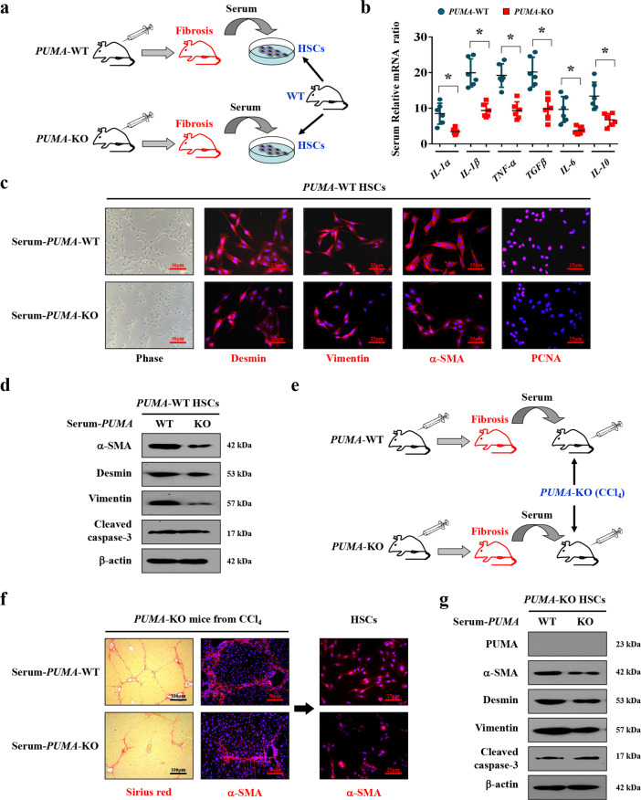
Fig. 9. NF-κBp65/PUMA-driven liver inflammation-induced HSCs activation and liver fibrosis.
a Schematic diagram of the serum test in vitro. b The genes expressions of the indicated inflammatory cytokines from CCl4-treated PUMA-WT or PUMA-KO mice were detected by quantitative reverse-transcription PCR. The expression of β-actin in each tissue was quantified as the internal control. n = 6 per group. *P < 0.05. c Representative images of the growth with activation of primary isolated HSCs following the treatment of the distinct serum extracted from CCl4-treated PUMA-WT or PUMA-KO mice. Nuclei (blue) were counterstained with DAPI. d Expressions of the related proteins of primary HSCs after serum treatment were detected by western blotting, revealing that PUMA-WT serum enhanced the activation of HSCs without affecting cell apoptosis. n = 6 per group. e Schematic diagram of the serum test in vivo. f Sirius red staining (red) and α-SMA staining (red) were examined in the liver tissues of PUMA-KO mice following the treatment of the serum extracted either from CCl4-treated PUMA-WT or PUMA-KO mice. The primary isolated HSCs were also analyzed by α-SMA staining (red). Nuclei (blue) were counterstained with DAPI. g The indicated proteins of the primary isolated HSCs from PUMA-KO mice after serum treatment were detected by western blotting, n = 6 per group.

