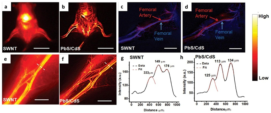
Figure 6.

Whole body and vascular imaging in NIR-II optical window by using SWNT and PbS/CdS NIR-II fluorophores. a, b) Whole body NIR-II fluorescence imaging after tail-vein injection of (a) SWNTs and (b) PbS/CdS. c, d) Unambiguous imaging of femoral artery and vein after tail-vein injection of (c) SWNTs and (d) PbS/CdS. e, f) High-magnification images of the hindlimb vasculature imaged by (e) SWNTs and (f) PbS/CdS. g, h) Representative cross-sectional fluorescence intensity profiles of (e) SWNTs and (f) PbS/CdS. e, f) Gaussian fits to the profiles are shown in red dashed curves. Scale bar: 2 cm (a, b), 5 mm (c, d), 2 mm (e, f). Reproduced with permission.[30] Copyright 2018, John Wiley and Sons.
