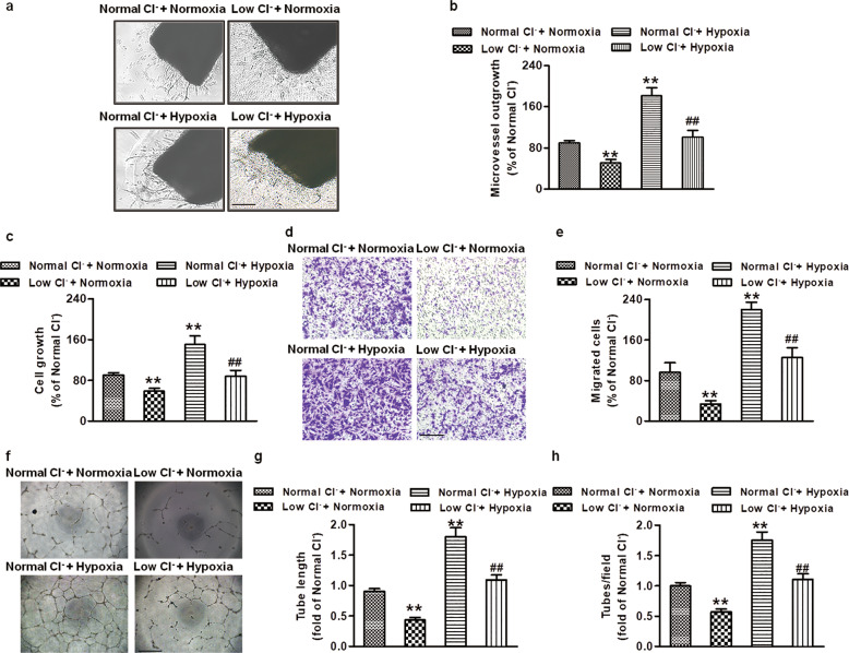
Fig. 2. Low Cl− inhibits hypoxia-induced angiogenesis in vitro.
a Representative phase-contrast micrographs of aortic rings cultured with normal Cl− or low Cl− medium for 4 days under normoxic (21% O2) or hypoxic (1% O2) conditions. Scale bar = 200 µm. b Quantification of vascular outgrowth assessed by Image-Pro Plus software. **P < 0.01 vs. normal Cl− + normoxia; ##P < 0.01 vs. normal Cl− + hypoxia, n = 5. c Cell proliferation of HUVECs was detected by CCK-8 assays. **P < 0.01 vs. normal Cl− + normoxia; ##P < 0.01 vs. normal Cl− + hypoxia, n = 6. d Representative images of the transwell assay to assess HUVEC migration stained by crystal violet under normoxic or hypoxic conditions. Scale bar = 100 µm. e The relative migrated cell numbers were quantified by measuring solubilized crystal violet. **P < 0.01 vs. normal Cl− + normoxia; ##P < 0.01 vs. normal Cl− + hypoxia, n = 6. f Representative pictures of HUVEC tube formation under normoxic or hypoxic conditions. Scale bar = 200 µm. Quantification of tube formation assessed by the number of tubes (g) and length of tubes (h). **P < 0.01 vs. normal Cl− + normoxia; ##P < 0.01 vs. normal Cl− + hypoxia, n = 6.

