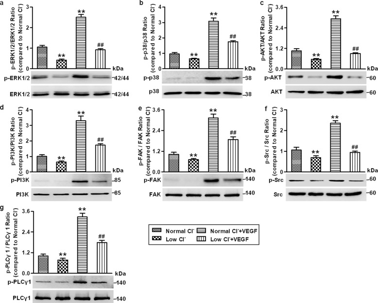
Fig. 7. Low Cl− medium blocks VEGFR2 downstream axis activation in HUVECs.
a–g The cells were cultured in normal Cl− or low Cl− medium in the presence or absence of VEGF (50 ng/mL) for 48 h. The phosphorylation of ERK1/2 (a), p38 (b), AKT (c), PI3K (d), FAK (e), Src (f), and PLCγ1 (g) was determined by Western blotting. GAPDH was used as a loading control. Densitometric analysis of the phosphorylated levels of the above proteins. **P < 0.01 vs. normal Cl−; ##P < 0.01 vs. normal Cl− + VEGF, n = 6.

