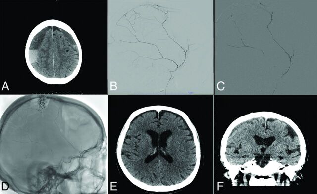
FIG 1.
Treatment course of a 77-year-old man with bilateral cSDH. Noncontrast axial head CT reveals bilateral subacute-on-chronic SDHs on admission (A). Diagnostic cerebral angiogram reveals robust filling of the MMA (B). The MMA was then embolized using n-BCA diluted with D5W (C). Postprocedural spin sequence reveals the glue cast left in the MMA (D). Noncontrast head CT at 3 months reveals significant resolution of the patient's SDHs (E and F).

