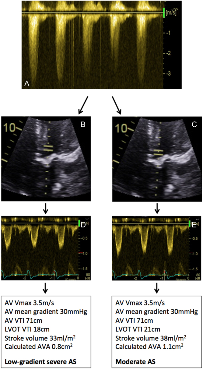
Figure 15.
Example of technical error resulting in low-gradient AS. A patient was noted to have calcified restricted aortic valve. The LVOTd is 2.0 cm (not shown). The LVOT cross-sectional area is 3.14 cm2. PEDOF values from the apical 5-chamber window demonstrate an AV Vmax 3.5 m/s; mean gradient 30 mmHg; AV VTI 71 cm (A). If the PW sample volume is placed as in image (B), corresponding Doppler traces are shown in (D). This leads to an estimated AVA of 0.8 cm2, consistent with low-gradient severe AS. In image (C), the PW sample volume has been moved closer to the valve, with corresponding Doppler trace in (E): note the amplitude of the traces is significantly larger. This leads to an estimated AVA of 1.1 cm2, consistent with moderate AS.

 This work is licensed under a