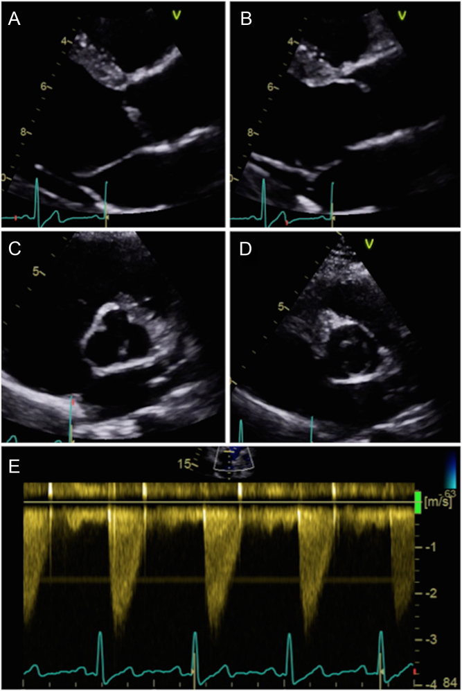
Figure 3.
Parasternal long-axis window of a unicuspid aortic valve during diastole (A) and systole (B). Note the marked ‘doming appearance’ during systole. Parasternal short-axis window during diastole (C) and systole (D): note how the orifice is eccentric. This valve has minimal calcification and retains mobility. Image (E) represents CW Doppler through the unicuspid valve. There is an obstruction to flow despite the lack of calcification.

 This work is licensed under a