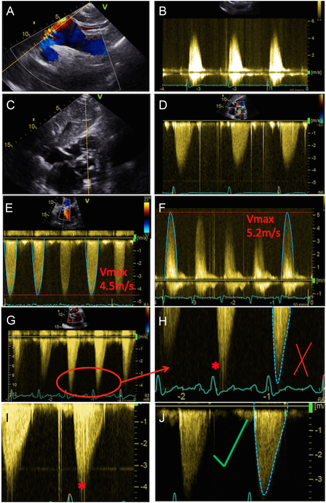
Figure 5.
Assessment of maximal AV velocity and mean gradient using CW Doppler. CW tracings should be obtained from multiple echocardiographic windows: suprasternal window (A), with the corresponding Doppler in (B); subcostal (C) and (D); apical 5-chamber (E); and stand-alone probe from the right parasternal window (F). Note that the AV Vmax in (E) represents a significant underestimation of maximal velocity when compared to that obtained using the standalone probe (F). Images (G) and (H) demonstrate spectral dispersion of the CW Doppler trace (marked with an asterisk). The mean gradient is obtained from tracing around the dense part of the CW Doppler curve. In images (I) and (J), the CW trace has been optimised: in (I) some spectral dispersion remains, but this has been completely eliminated in (J), resulting in an ideal CW trace.

 This work is licensed under a