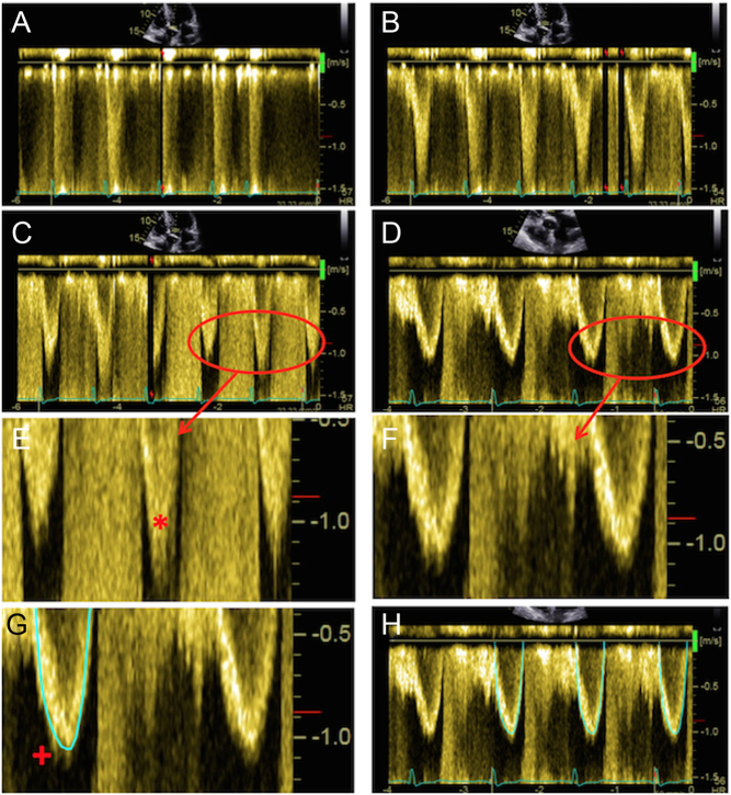
Figure 7.
Optimal assessment of the LVOT Doppler. The PW Doppler sample volume should initially be placed on the aortic valve (A), which will usually result in aliasing. It should then be slowly moved apically (i.e. away from the valve; images B and C). In (C) there is a wide area of density at the apex of the trace, evident in the zoomed image (E; marked with asterisk). This represents blood flow within the zone of acceleration immediately proximal to the valve, and should not be included, as it would result in an overestimation of LV stroke volume and an underestimation of AS severity. In (D), (F), (G) and (H) the trace has been optimised further and depicts ideal assessment of LVOT Doppler. When tracing the curve, any spectral dispersion should be ignored (marked in (G) with a +). Three traces should be obtained and averaged for use in the continuity equation, with sweep speed set between 50 and100 mm/s (H).

 This work is licensed under a