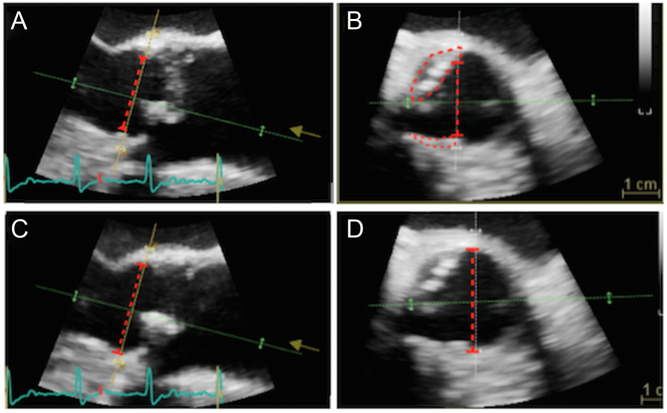
Figure 8.
Eccentric calcification can lead to important underestimation of LVOT diameter, here demonstrated with 3D imaging. (A) Depicts a long-axis view obtained during TOE with LVOT diameter marked (red dotted line). Picture (B) is an image of the LVOT taken at an orthogonal plane to image (A). Note that the LVOT diameter is an underestimate owing to two areas of eccentric calcification (marked). Adjustment of the imaging plane can remove calcification from the image (C), with the optimal LVOT diameter now shown (red dotted line in images C and D).

 This work is licensed under a