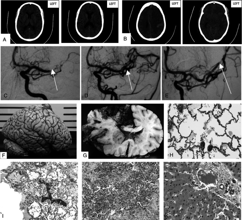
FIGURE 1.

A, Initial precontrast CT brain scan showing loss of definition of the lateral left lentiform nucleus and insular ribbon with mild sulcal effacement and loss of gray-white matter differentiation in the high MCA territory. B, Follow-up uncontrasted CT brain scan showing malignant left hemisphere infarction with small parenchymal hemorrhage and associated mass effect. C, Initial angiogram of left MCA with arrow indicating site of occlusion. D, Angiogram post–pump aspiration of left MCA with arrow showing restoration of flow. E, Angiogram of the left MCA showing persistent occlusion of the angular branch. F, External surface of fixed brain demonstrating swelling. G, Coronal section of fixed brain with left AMA and MCA territory hemorrhagic infarction and left-to-right midline shift. H, Pulmonary blood vessels with fibrin thrombi (hematoxylin-eosin stain, original magnification ×200). I, Lung parenchyma with intravascular fibrin thrombi, scattered interstitial lymphocytes, and intra-alveolar macrophages (hematoxylin-eosin stain, original magnification ×100). J, Focal pulmonary fibrosis with scattered lymphocytes (hematoxylin-eosin stain, original magnification ×100). K, Hepatic portal tract with fibrin thrombus in the venule and adjacent glycogenated hepatocyte nuclei (hematoxylin-eosin stain, original magnification ×400). L, Spleen demonstrating hemosiderin deposition (Perls Prussian blue stain, original magnification ×100). M, Left ACA territory infarct with intravascular fibrin thrombus (Martius scarlet blue stain, original magnification ×200). N, Left ACA territory infarct with fibrin thrombus in blood vessel, surrounded by mixed inflammatory cells and associated spongiosis (hematoxylin-eosin stain, original magnification ×400). O, Left MCA territory hemorrhagic infarct demonstrating red neurons, neutrophils, and macrophages (hematoxylin-eosin stain, original magnification ×200). P, Left MCA territory infarct with red neurons (hematoxylin-eosin stain, original magnification ×400). Q, Pontine microinfarct with axonal spheroids and spongiosis (hematoxylin-eosin stain, original magnification ×400). R, Pontine microinfarct with red neurons and spongiosis (hematoxylin-eosin stain, original magnification ×200). S, Pontine blood vessel fibrin thrombus (hematoxylin-eosin stain, original magnification ×200). T, Right MCA microinfarct with spongiosis, axonal spheroids, and perivascular inflammatory cells (hematoxylin-eosin stain, original magnification ×200). U, Right ACA territory blood vessel at leptomeninges with surrounding mixed inflammatory cells (hematoxylin-eosin stain, original magnification ×400; [I, J] hematoxylin-eosin stain, original magnification ×100; [H, O, R, S, T] hematoxylin-eosin stain, original magnification ×200; [K, N, P, Q, U] hematoxylin-eosin stain, original magnification ×400; [L] Perls Prussian blue stain, original magnification ×100; [M] Martius scarlet blue stain, original magnification ×200).
