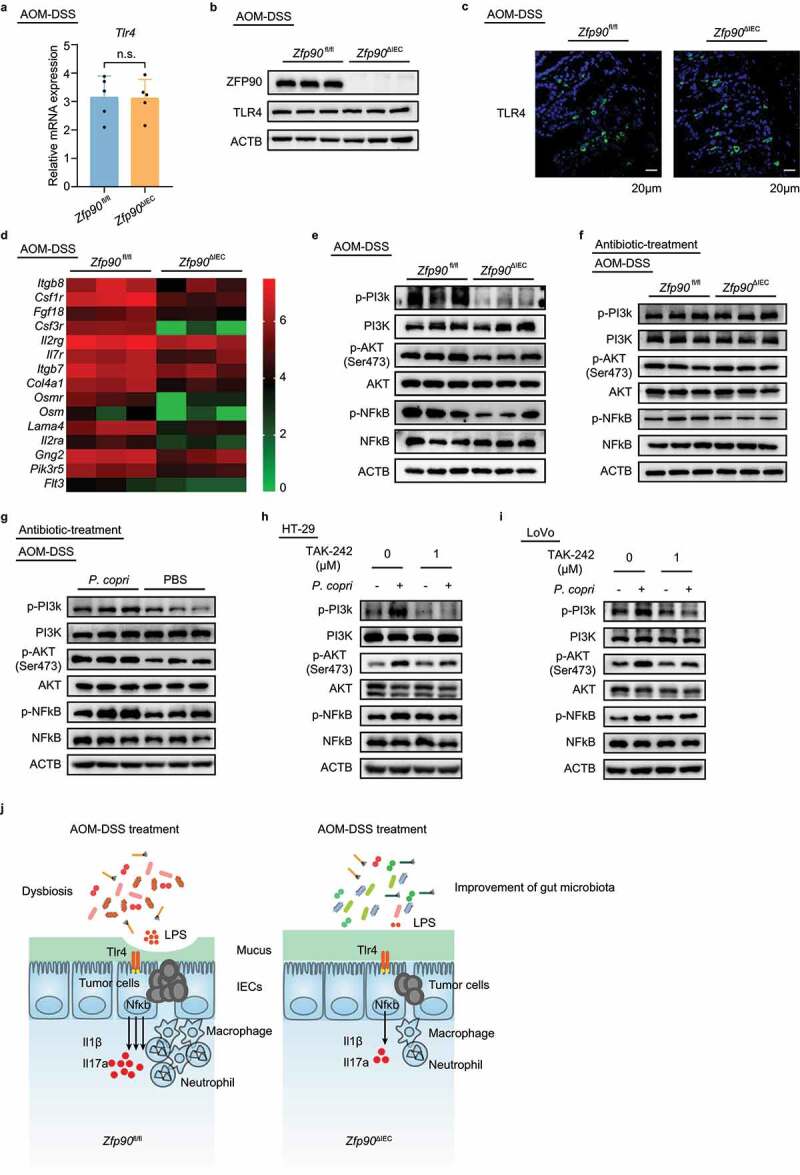
Figure 5.

Decreased TLR4-dependent PI3K/AKT/NF-κB signaling in colon tissue of Zfp90ΔIEC mice
a Real-time PCR was performed to determine the mRNA expression of Tlr4 in AOM-DSS-treated Zfp90fl/fl and Zfp90ΔIEC mice (n = 5 per group). b Representative data showing protein level of TLR4 in AOM-DSS-treated Zfp90fl/fl and Zfp90ΔIEC mice. c Representative immunofluorescence staining of the TLR4 protein in tumor tissues from AOM-DSS-treated Zfp90fl/fl and Zfp90ΔIEC mice. Sections were stained with DAPI (blue) and TLR4 (green). d Heatmap representing log2-normalized count of the PI3K-AKT pathway expression on the colonic epithelium from Zfp90fl/fl and Zfp90ΔIEC mice (n = 3 per group). e Representative data showing the activity of the PI3K-AKT-NFκB pathway in AOM-DSS-treated Zfp90fl/fl and Zfp90ΔIEC mice. f Representative data showing the activity of the PI3K-AKT-NFκB pathway in antibiotic-treated Zfp90fl/fl and Zfp90ΔIEC mice after AOM-DSS treatment. g Representative data showing the activity of the PI3K-AKT-NFκB pathway in PBS- and P. copri-treated WT mice after AOM-DSS treatment. h, i HT-29 (h) and LoVo (i) cell lines were pretreated with TLR4 inhibitor (TAK-242) 30 minutes before co-culturing with P. copri. Representative data showing the effect of TLR4 inhibitor on the activation of the PI3K-AKT-NFκB pathway. j Model for the P. copri-defined microbiota-mediated colorectal tumorigenesis. Data with error bars represent the mean ± SD. Each panel is a representative experiment of at least three independent biological replicates. Statistical analysis was performed using nonpaired two-tailed t-test.
